
Figure 5. The design and application of the repressilator.
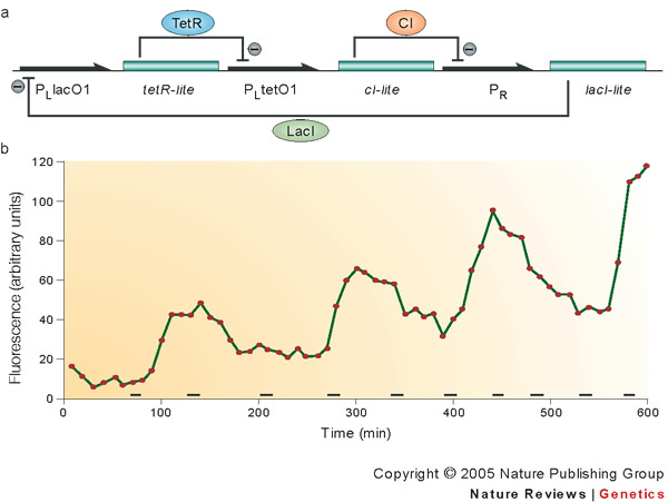
a | Schematic showing the regulation pattern that forms the basis of a repressilator. Three gene–promoter pairs are arranged so that the product derived from the expression of the gene following a promoter is a repressor for the next promoter in the cycle. Black connecting lines show that promoter PLlacO1 controls the transcription of the gene tetR-lite, the tetracycline repressor protein TetR represses PLtetO1, which is the next promoter in the sequence. PLtetO1 in turn controls the transcription of cI-lite, and the protein CI represses the promoter PR. Finally, PR controls the expression of lacI-lite, and the lactose repressor protein LacI represses PLlacO1, completing the cycle. The suffix '-lite' refers to the presence of tags that increase the degradation rate of the proteins. b | The luminescence pattern of a reporter plasmid that carries GFP under the transcriptional control of the PLtetO1 promoter, when the reporter construct is transferred to an Escherichia coli in the presence of the repressilator. As the experimental trace shows, the oscillation of the TetR repressor expressed from the repressilator results in the time dependent oscillation of GFP expression. Bars at the bottom of the diagram show the timing of cell division events. The period of the oscillations is longer than the cell division time, and the cycle of oscillations continues in the subsequent generations. Adapted, with permission, from Nature Ref. 16 © (2000) Macmillan Magazines Ltd.