
Figure 5. Adaptation of blood flow to ventilation in the pulmonary circulation.
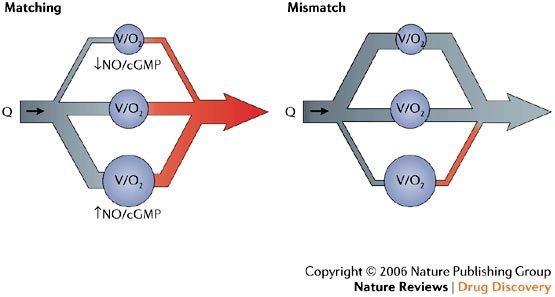
Blood flow (Q) in the pulmonary circulation must, ideally, be directed to well-ventilated areas (symbolized by big V (ventilation) and O2 (oxygenation) in the largest alveolus (blue circle at bottom of figure)) to ensure optimized gas exchange ('matching'), whereas only a small amount of blood should flow through areas of minor or no ventilation (midsize and small alveolus, respectively) (left panel). Lung vessel dilatation is mainly regulated by the compartmentalized production of nitric oxide (NO) and subsequent intracellular cGMP formation, where alveolar distension and oxygenation represent the most potent stimuli for this local NO release. Similarly, less NO/cGMP is produced in non-ventilated areas of the lung, resulting in hypoxic vasoconstriction (the so called von Euler–Liljestrand mechanism). During application of non-selective vasodilators and/or under disease conditions (for example, chronic obstructive lung disease, lung fibrosis, sepsis or acute respiratory distress syndrome), vasodilatation is induced in poorly or non-ventilated areas of the lung resulting in venous admixture and worsening of gas-exchange ('mismatch', right panel). There is strong evidence that oral sildenafil preferentially dilates vessels in well-ventilated areas of the lung, thereby both reducing overall vascular resistance and improving overall oxygenation ('re-matching' drug)143,144.