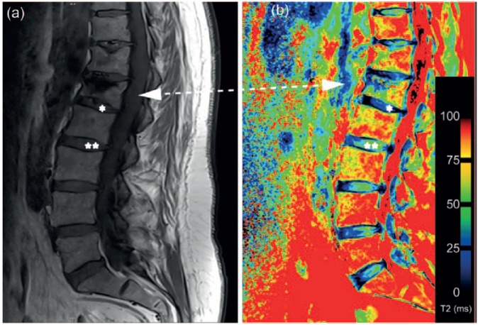
Figure 2.
(a) Morphological magnetic resonance image of the lumbar spine. Vertebral body after kyphoplasty (L1) marked by arrow. Intervertebral discs adjacent to vertebral body after kyphoplasty marked with an asterisk (*), intervertebral discs nonadjacent to vertebral body after kyphoplasty marked with double asterisks (**). (b) T2 map with clearly visible differences between intervertebral discs adjacent (*) and nonadjacent (**) to vertebral bodies after kyphoplasty.

