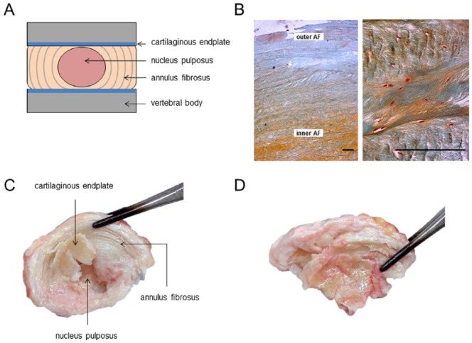
Figure 1.
Intervertebral disc morphology and degenerative disc disease. (A) Schematic representation of an intervertebral disc. The IVD comprises 3 distinctive tissues: a central gelatinous nucleus pulposus (NP), surrounded by the lamellar annulus fibrosus (AF). The AF consists of multiple, concentrically arranged lamellae in which cells and ECM components interact to provide function and structure. In the AF, two distinct zones are distinguished primarily based on their collagen/proteoglycan content: the inner and outer AF. The NP and AF are flanked at each side by cartilaginous endplates (CEP), which mediate the contact between the IVD body and vertebra. (B) Sections of a healthy AF (15-year-old male donor) stained with Safranin O and Fast green. The outer AF predominantly contains collagens while the inner AF contains proteoglycans. The characteristic lamellar structure is clearly visible; bar represents 200 µm. (C) A nondegenerate L1/L2 intervertebral disc of 63-year-old male donor. The lamellar annulus fibrosus is clearly recognizable. The central nucleus pulposus displays no aberrant morphology; the cartilaginous endplate is relatively thin. (D) The L4/L5 intervertebral disc of the same donor. Degenerative changes cause apparent loss of lamellar structure in the AF. The NP is no longer distinguishable from the AF and additional ossification is found near the CEP (For interpretation of the references to colours in this figure legend, refer to the online version of this article).

