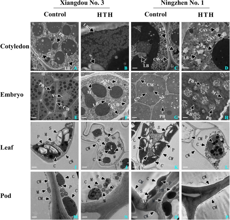
Fig. 2.

Transmission electron micrographs of soybean cvs. Xiangdou No. 3 and Ningzhen No. 1. Microstructure changes in cotyledon (A, B, C, D), embryo (E, F, G, H), leaf (I, J, K, L) and pod (M, N, O, P) of both the cultivars under the HTH stress (96 h) and control (96 h). A, C, I, K, 5.0 μm; E, G, J, L, 10.0 μm; B, D, 1.0 μm; F, H, M-P, 2.0 μm; C, chloroplast; CAV, cavitation; CW, cell wall; CM, cell membrane; CN, cell nucleus; LB, lipid body; M, mitochondrion; P, phagosome; PB, protein body; S, starch grain
