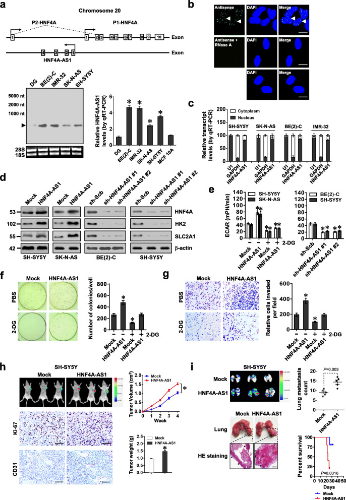
Fig. 2.
HNF4A-AS1 promotes aerobic glycolysis and NB progression. a Schematic illustration indicating genomic location of HNF4A-AS1 and HNF4A. Northern blot using a 254-bp specific probe and real-time qRT-PCR (normalized to β-actin, n = 5) showing the endogenous existence of HNF4A-AS1 transcript in normal dorsal root ganglia (DG), NB cell lines, and MCF 10A cells. b RNA-FISH using a 254-bp antisense probe showing localization (arrowheads) of HNF4A-AS1 in the nuclei (DAPI staining) of BE(2)-C cells, with sense probe and RNase A (20 μg) treatment as negative controls. Scale bars, 10 μm. c Real-time qRT-PCR (normalized to β-actin, n = 4) revealing the enrichment of HNF4A-AS1 in the cytoplasm and nuclei of NB cells. d Western blot assay indicating the expression of HNF4A and glycolytic genes in SH-SY5Y, SK-N-AS, and BE(2)-C cells stably transfected with empty vector (mock), HNF4A-AS1, scramble shRNA (sh-Scb), or sh-HNF4A-AS1. e–g ECAR bars (e), soft agar (f), and matrigel invasion (g) assays showing glycolysis, anchor-independent growth, and invasion of NB cells stably transfected with mock, HNF4A-AS1, sh-Scb, or sh-HNF4A-AS1, and those treated with 2-DG (10 mmol·L−1, n = 4). h Representative images, in vivo growth curve, Ki-67 and CD31 immunostaining, and weight at the end points of subcutaneous xenograft tumors formed by SH-SY5Y cells stably transfected with mock or HNF4A-AS1 in nude mice (n = 5 per group). Scale bar, 100 μm. i In vivo imaging, representative images and metastatic counts of lungs, and Kaplan-Meier curves of nude mice (n = 5 per group) treated with tail vein injection of SH-SY5Y cells stably transfected with mock or HNF4A-AS1. Scale bar, 100 μm. ANOVA and Student’s t test compared the difference in a and e–i. Log-rank test for survival comparison in i. *P < 0.05 vs. DG, mock, or sh-Scb. Data are shown as mean ± s.e.m. (error bars) and representative of three independent experiments in a–g

