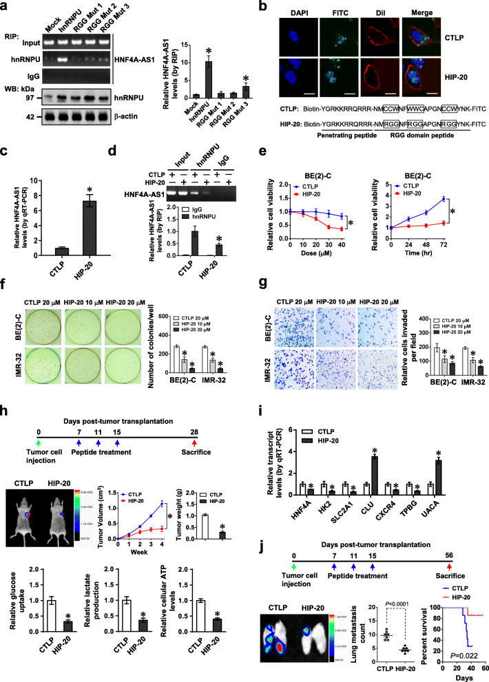
Fig. 5.
Therapeutic blocking HNF4A-AS1-hnRNPU interaction inhibits aerobic glycolysis and NB progression. a RIP and real-time qRT-PCR assays showing the binding of hnRNPU to HNF4A-AS1 in BE(2)-C cells stably transfected with wild-type or RGG mutant form of hnRNPU. b Representative images indicating the distribution of FITC-labeled RGG mutant control (CTLP) or hnRNPU inhibitory peptide (HIP-20, 20 μmol·L−1) in BE(2)-C cells, with nuclei and cellular membranes staining with DAPI or Dil. Scale bar, 10 μm. c and d Real-time qRT-PCR (c, normalized to β-actin, n = 4) and RIP (d) assays revealing HNF4A-AS1 pulled down by biotin-labeled CTLP or HIP-20 (20 μmol·L−1), and interaction of HNF4A-AS1 with hnRNPU in BE(2)-C cells treated with CTLP or HIP-20 (20 μmol·L−1). e–g MTT colorimetric (e), soft agar (f), and matrigel invasion (g) assays indicating the viability, anchorage-independent growth, and invasion of NB cells treated with different doses of CTLP or HIP-20 for 24 h, or 20 μmol·L−1 peptides for time points as indicated (n = 5). h and i Representative images (h, left middle panel), in vivo growth curve (h, left middle panel), tumor weight (h, right middle panel), glucose uptake, lactate production, and ATP levels (h, lower panel), and HNF4A-AS1 downstream gene expression (i, normalized to β-actin) of BE(2)-C-formed subcutaneous xenograft tumors (n = 5 per group) that were treated with intravenous injection of CTLP or HIP-20 (5 mg·kg−1) as indicated (h, upper panel). j Representative images and metastatic counts of lungs and Kaplan-Meier curves (lower panel) of nude mice (n = 5 per group) treated with tail vein injection of BE(2)-C cells and CTLP or HIP-20 (5 mg·kg−1) as indicated (upper panel). ANOVA and Student’s t test compared the difference in a and c–j. Log-rank test for survival comparison in j. *P < 0.01 vs. mock or CTLP. Data are shown as mean ± s.e.m. (error bars) and representative of three independent experiments in a–g

