Fig. 1.
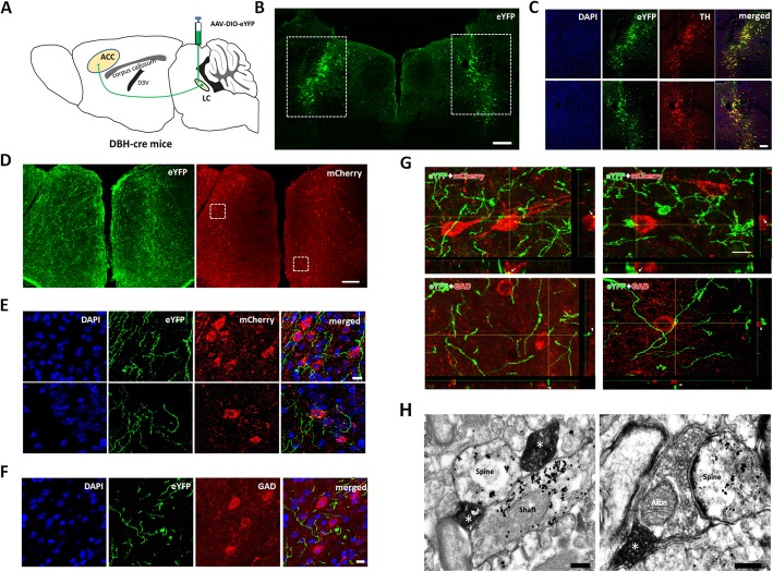
Noradrenergic fibers from the LC project to the postsynaptic pyramidal cells in the ACC. a Diagram of AAV-DIO-eYFP injection and anterograde tracing strategy in the DBH-cre mice. b The AAV-DIO-eYFP injection sites in the LC. c AAV infected neurons in the rectangle areas of b are dual labeled with TH- immunopositivities. d Distribution of LC-ACC projecting NAergic fibers showing eYFP and the pyramidal cells infected by AAV-CaMKIIα-mCherry in the ACC. e Enlarged figures in the rectangle areas in d showing LC-ACC projecting fibers (eYFP) made close connections with pyramidal cells (mcherry). f LC-ACC projecting NAergic fibers (eYFP) does not make close connection with GAD immunoreactive GABAergic neurons. g The 3D view of the close connections between eYFP labeled LC-ACC fibers and mcherry-labeled pyramidal cells or GAD-immunoreactive GABAergic neurons. h Two LC-ACC NAergic (eYFP immunoreactive with DAB) axon terminals (*) simultaneously make synapses with a dendritic spine and a shaft of a pyramidal cell (mcherry immunoreactive with nanogold) (left panel); One LC-ACC NAergic terminal (*) makes a synapse with a glutamatergic axon terminal, which in sequence makes a synapse with a spine of a pyramidal cell (right panel). Bars equals to 200 nm in h, 10 μm in e, f and g, 100 μm in C and 200 μm in b and d

