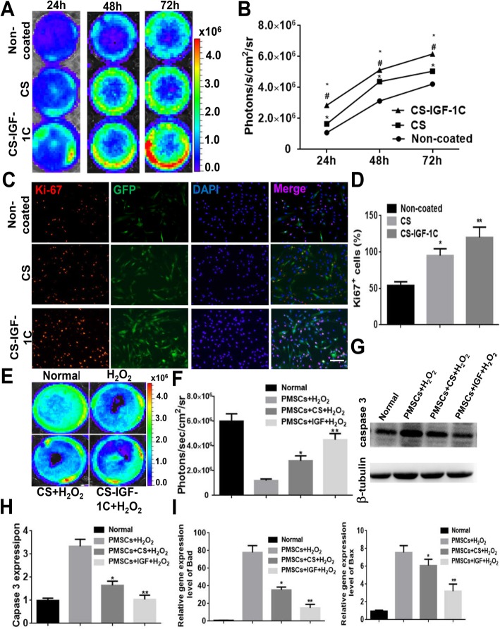
Fig. 2.
Proliferative and protective effects of CS-IGF-1C hydrogel in vitro. a BLI exhibited that CS-IGF-1C hydrogel enhanced the proliferation of hP-MSCs. b Quantitative analysis of BLI signals. The signal activity expressed as photons/second/cm2/steradian. *P < 0.05 versus non-coated; #P < 0.05 versus CS. c Representative images showed the proliferation (Ki-67, red) of hP-MSCs (GFP, green) incubated with CS or CS-IGF-1C hydrogel for 24 h. The bar represented 100 μm. d Quantification of the proliferation index of hP-MSCs performed by CS or CS-IGF-1C hydrogel. *P < 0.05 versus non-coated; **P < 0.01 versus non-coated. e Anti-apoptotic effects of CS-IGF-1C hydrogel. BLI revealed that CS-IGF-1C hydrogel protected hP-MSCs under oxidative condition. f Quantification of BLI signals displayed a significant amelioration when cultured on CS-IGF-1C hydrogel-coated plates after H2O2 treatment. *P < 0.05 versus non-coated; **P < 0.01 versus non-coated. g Caspase-3 and Tubulin expressions were detected by Western blots. h Quantification of Caspase-3 expression. i Real-time quantitative PCR analysis of apoptosis-related gene expression of hP-MSCs after treated with H2O2 for 4 h. All data were shown as means ± SEM in triplicate assays. *P < 0.05 versus PMSCs + H2O2; **P < 0.01 versus PMSCs + H2O2

