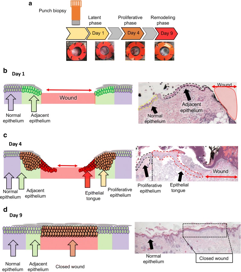
Fig. 1.
Epidermal wound and histological analysis: a Schematic representation of the experiment timeline showing the punch biopsy tool to create a 5 mm wound on the skin. Silicone splint was attached to the back of the mice to prevent wound contraction. Tissue samples were collected 1, 4, and 9 days after wounding (n = 8 mice per time point). b Schematic representation and H&E staining of wounds collected on day 1. Note the presence of thin adjacent epithelium next to the open wound of mice. c Schematic representation and H&E staining of wound collected on day 4. Note the formation of an epithelial tongue migrating underneath the wound scab, and the presence of a thick proliferative epithelium constituted by proliferative epithelial cells. d Schematic representation and H&E staining of wound area collected on day 9. The wound is closed (reepithelization) by day 9. The repaired epidermis presents a thick layer of epithelial cells that covers the wound (box) when compared to the normal epidermis (arrow)

