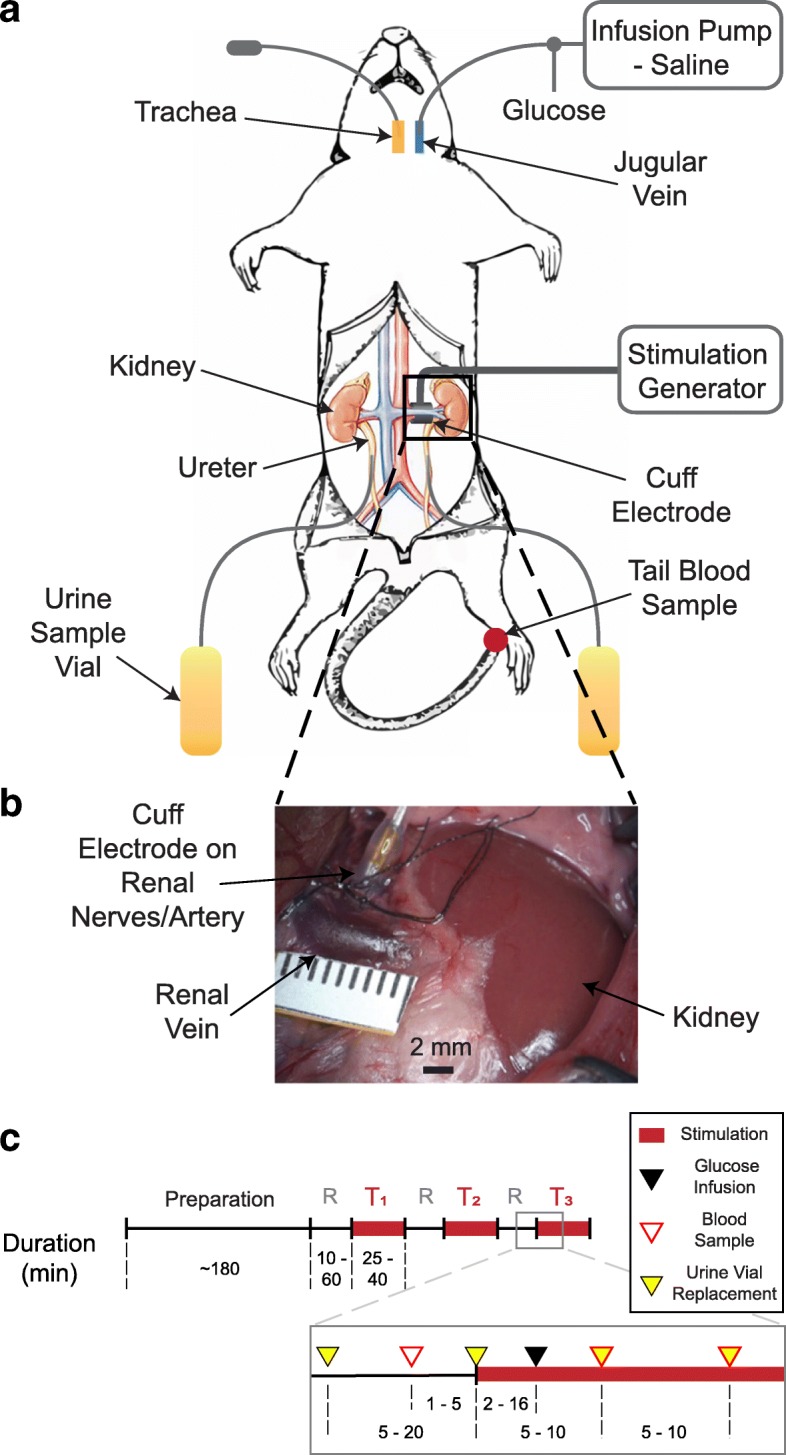
Fig. 1.

Experimental setup diagram and protocol timeline. a Experimental setup: Jugular vein was cannulated for saline and glucose infusion. Nerve cuff electrode was placed on renal nerves of the left kidney and connected to a stimulation generator. Ureters were cannulated bilaterally, and urine samples were collected in sampling vials. b Nerve cuff electrode was placed around the renal artery, encapsulating the renal nerve branches that run along the renal artery. c Timeline for experimental protocol: Each experiment consisted of 1–3 stimulation trials (T1-T3), with a rest period (R) before each trial. A glucose bolus was infused in each trial. Blood glucose measurements and urine samples were obtained periodically throughout the trials
