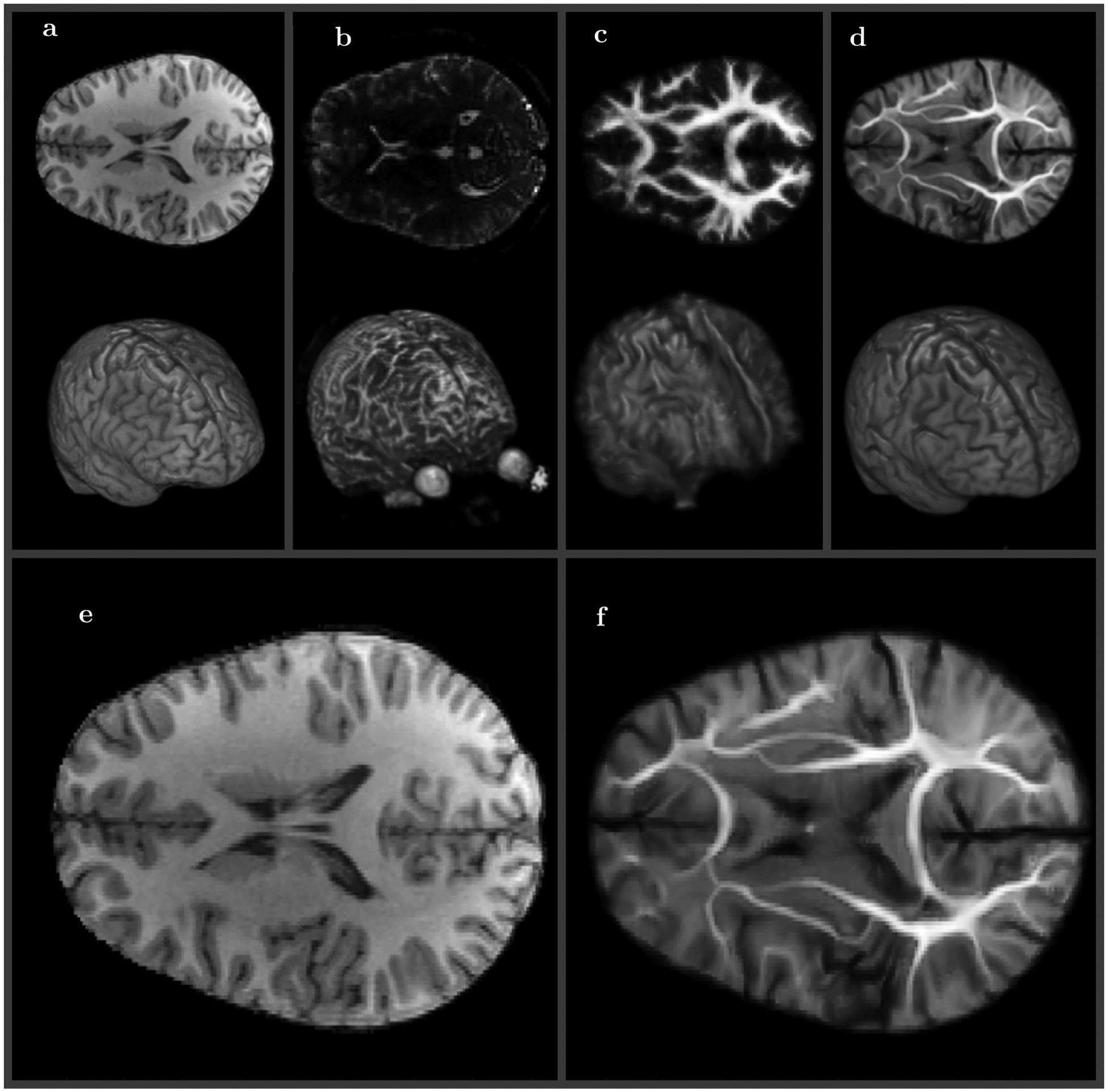
FIGURE 4.

Medium resolution (100 × 100 × 72) diffusion weighted (DWI) volume registration to high resolution (168 × 256 × 256) T1 reference. A, Reference T1 MRI image (2D center slice top and 3D view bottom), B, DWI b0 MRI image, C, equilibrium probability DWI image (same resolution as b0 image), D, DWI image SWD preconditioned and registered to T1 image (same resolution as T1 image). Side by side comparison of reference E, and symplectomorphic registration of DWI volume F (enlarged versions of A and D)
