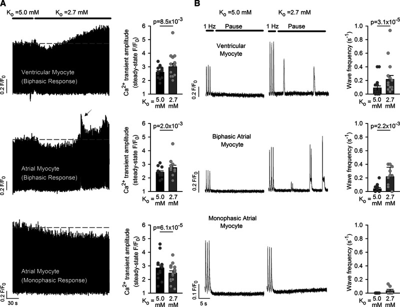
Figure 1.

Hypokalemia promotes a steady-state increase in Ca2+ transients and Ca2+ waves in ventricular myocytes and a subpopulation of atrial myocytes. A, In field-stimulated ventricular cells, rapidly lowering [K+]o from 5.0 to 2.7 mmol/L produced an initial depression of Ca2+ transient magnitude. A secondary rising phase followed which ultimately yielded larger Ca2+ transients compared with control conditions (right, n=15 cells, 8 hearts). A similar biphasic response to hypokalemia was observed in some atrial cardiomyocytes (13 of 31 cells, 10 hearts), with associated over-activity (arrow). Other atrial cells exhibited only a monophasic reduction in Ca2+ transient amplitude. B, Ca2+ waves were assessed during pauses in the electrical stimulus. Ventricular cells and those atrial cells which exhibited a biphasic response demonstrated an increased frequency of Ca2+ waves during hypokalemia. For Ca2+ wave measurements, ncells=17, 7, 12; nhearts=10, 5, 11 in ventricular, biphasic atrial, and monophasic atrial populations. Statistics: Wilcoxon signed-rank test.
