
Figure 2.
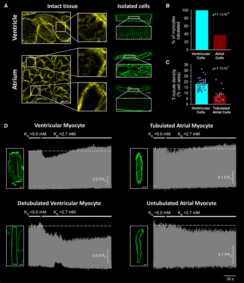
The biphasic Ca2+ transient response to hypokalemia requires t-tubules. A, T-tubule staining in intact tissue and isolated cells (caveolin-3 and di-8-ANEPPS, respectively) revealed a dense and well-organized t-tubule network in ventricular cells (scale bars=10 µm). T-tubules were only observed in approximately one-third of atrial cells (B, 21 of 56 cells, 4 hearts), and when present, these tubules were less well organized and at lower density (C) than in ventricular cells (40 cells, 3 hearts). D, Paired imaging of t-tubules and Ca2+ revealed that only tubulated atrial cells exhibited a biphasic response to hypokalemia (n=12 cells, 10 hearts), while untubulated cells exhibited a monophasic decline in Ca2+ transient amplitude (n=7 cells, 5 hearts). A similar monophasic decline in Ca2+ transients was reproduced in experimentally detubulated ventricular myocytes (n=10 cells, 3 hearts). Statistics: (B): z-test; (C): Mann-Whitney rank-sum test.