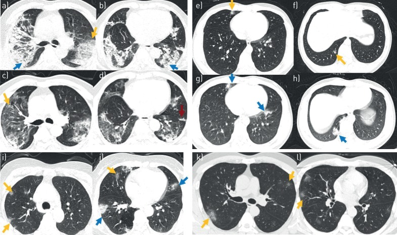
FIGURE 1.
High-resolution computed tomography (HRCT) manifestations of selected four cases with coronavirus disease 2019 (COVID-19). Case 1: 57-year-old male patient. HRCT scan was performed on day of admission (8 days after the onset of illness), showing a) a combined pattern of consolidations (blue arrow) and ground-glass opacities (GGOs) (yellow arrow) with air bronchograms in both lobes, and b) areas of consolidation (blue arrow) in both lower lobes. Follow-up HRCT scan performed 10 days after admission, showing c, d) remission of abnormalities, with reduced extent and density of airspace opacification, GGOs (yellow arrows) and fibrosis (red arrow). Case 2: 23-year-old male patient. HRCT scan was performed on day of admission (16 h after the onset of illness), showing e, f) a subpleural distribution of GGOs (yellow arrows) in the middle and lower right lobe. Follow-up HRCT scans performed 3 days after admission show g, h) larger areas of mixed consolidations and GGOs (blue arrows) in the upper right lobe and both lower lobes. Case 3: 50-year-old female. HRCT scan was performed on the day of admission (4 days after the onset of illness), showing i, j) a subpleural distribution of GGOs (yellow arrows) and consolidations (blue arrows) in both lobes. Case 4: 43-year-old female. HRCT scan was performed on day of admission (3 days after the onset of illness), showing k, l) a subpleural distribution of GGOs (yellow arrow) in both lobes.

