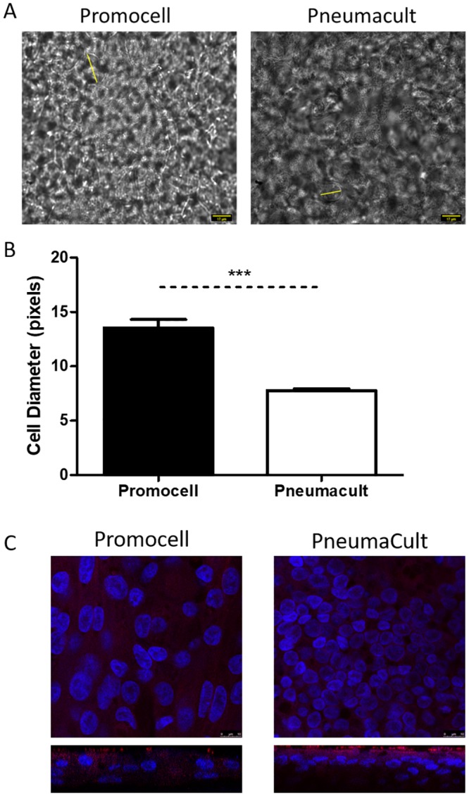
Fig 2. Primary paediatric nasal epithelial cells (n = 3 donors) seeded at 5x10⁴ per Transwell were differentiated and maintained using either Promocell or Pneumacult medium.

Cultures were fixed using 4% paraformaldehyde (PFA) on day 25 post air-liquid interface (ALI) initiation. Images were captured by DIC microscopy at x60 magnification and imageJ was used to determine the diameter of cells (yellow lines) (A). Graphical representation of the average cell diameter in pixels(B). Statistical significance was determined using unpaired t-tests. *** = p<0.001. Cultures were stained for beta-tubulin (red) and DAPI (blue). Z-stacks were obtained using a confocal microscope at x100 magnification (Leica SP5) (C).
