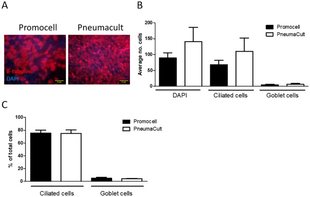
Fig 3. WD-PNEC cultures (n = 3 donors) with an initial seeding density of 5x10⁴ per Transwell were differentiated in Promocell or Pneumacult medium.
After 21 days cultures were fixed in 4% paraformaldehyde and stained for β-tubulin, a ciliated cell marker; Muc5ac, a goblet cell marker and counterstained for DAPI. Representative images of β-tubulin staining (A). The average number of total, ciliated and goblet cells from 5 fields of view per donor was calculated (B). The percentage of ciliated cells and goblet cells in the culture was calculated (C). Images were acquired using a Nikon Eclipse 90i at x60 magnification.

