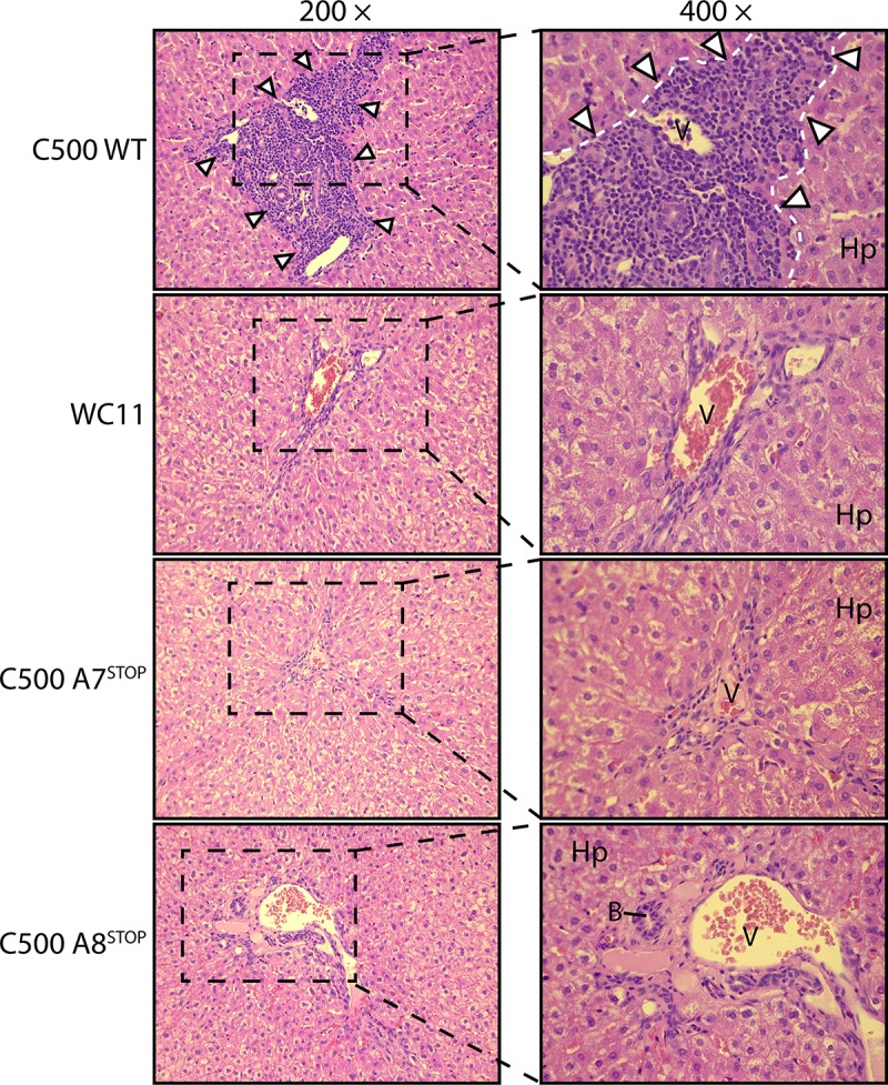
Fig 7. Histopathological characterization of MCF lesions.

Liver sections of one rabbit representative of each group are shown. White arrowheads indicate typical infiltrations of lymphoblastoid cells. Abbreviations: B, small bile ducts; Hp, hepatocytes; V, portal veins, Equivalent results were obtained in two independent experiments. Original magnifications are indicated.
