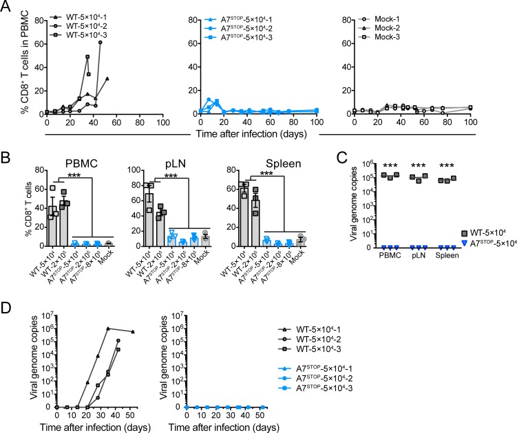
Fig 9. Lack of A7 results in the absence of expansion of CD8+ T lymphocytes and viral persistence in peripheral blood after intranasal infection.
Rabbits were infected by intranasal inoculation of different doses (5×10⁴, 2×10⁵ or 8×10⁵ PFU per rabbit) of C500 BAC− WT and BAC− A7STOP-207. (A) Percentages of CD8+ T cells in PBMCs analyzed by flow cytometry at regular intervals throughout the experiment. Data are plotted for individual rabbits. (B) Percentages of CD8+ T cells in PBMCs, popliteal lymph nodes (pLN) and spleen at the time of euthanasia, as analyzed by flow cytometry. Data show mean ± SEM with data plotted for each individual rabbit. (C) qPCR of viral genome copies in PBMCs over time. (D) qPCR of viral genome copies in PBMCs, pLN and spleen at the time of euthanasia. Real-time PCR quantification was normalized on 105 copies of the cellular β-globin gene sequence. Data are plotted as individual measurements. Bars show mean values. One-way ANOVA with Bonferroni’s post-hoc test was used to identify significant differences (***P<0.001).

