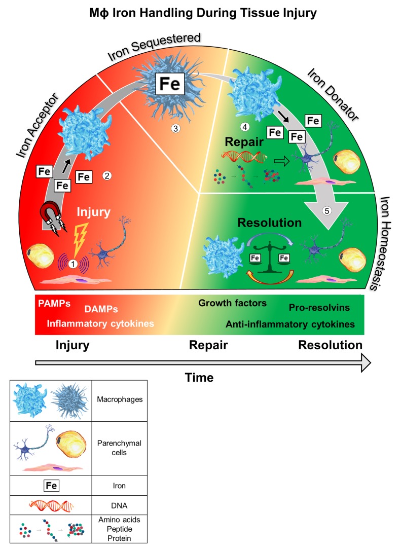
Figure 2. Proposed model by which resident iron-handling Mɸs regulate tissue homeostasis during an insult.
(1) Parenchymal cell injury initiates intracellular transduction signals that propagate transcriptional and nontranscriptional stress signals. (2) Local tissue-resident Mɸs are recruited to the injured site. Since injured cells are more susceptible to iron-induced oxidative damage, Mɸs sequester extracellular iron to decrease iron uptake in the injured parenchymal cell. LIP has been reported to increase in response to cell injury, which may promote iron efflux and Mɸ uptake. (3) A latency period follows, in which the Mɸ retains sequestered iron, allowing for parenchymal cell restoration. This phase of Mɸ iron retention may be aided by release of inflammatory cytokines and/or IFN responses that favor an iron-loaded Mɸ (i.e., suppression of Fpn-mediated iron export). Parenchymal cell repair requires iron for processes such as DNA synthesis. This utilization of parenchymal intracellular iron depletes ferritin stores. (4) The Mɸ mobilizes and relinquishes iron for parenchymal cell repair. (5) An undefined termination signal communicates cell resolution and the Mɸ regresses from the site, completing the homeostatic circuit and maintaining local iron balance.

