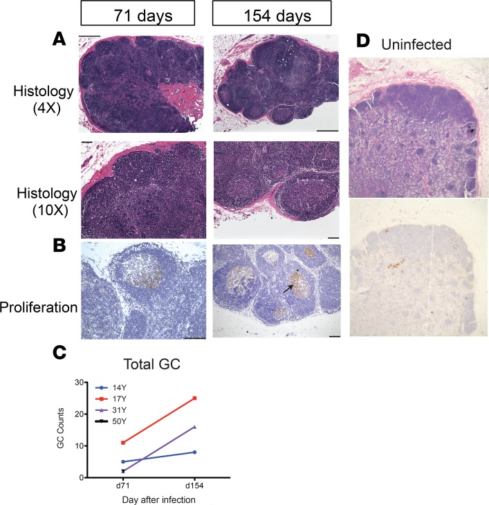
Figure 6. Lymph node germinal center histopathology.
Inguinal lymph node biopsies from uninfected macaques and measles virus–infected (MeV-infected) macaques 71 (n = 4) and 154 (n = 3) days after infection. Tissues were fixed, embedded in paraffin, sectioned, and stained with H&E (A and D) and for expression of Ki-67 indicative of cell proliferation (black arrow; B). Representative sections from 14Y (A) and 31Y (B) and an uninfected control macaque (magnification: ×4 objective; ×10 eyepiece) (D) are shown. Counts of germinal centers /lymph node section are indicated for 4 MeV-infected macaques from day 71 and 3 MeV-infected macaques from day 154 (C). 50Y data for d154 were not available and the d71 count is superimposed on data from 31Y. Scale bars: 2 μm (A: top panels; B: left panel) 100 μm (A: bottom panels; B: right panel)

