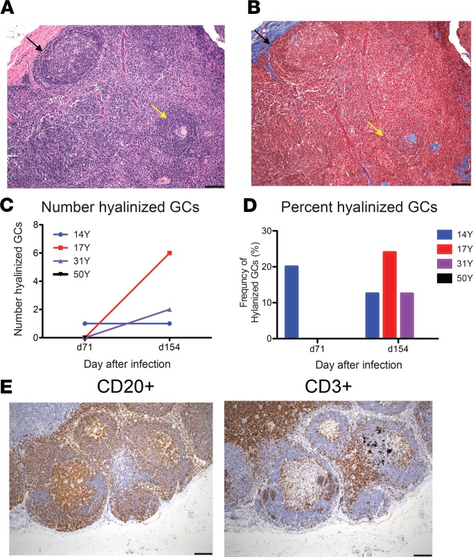
Figure 7. Germinal center hyalinization.
Sequential sections from inguinal lymph node biopsies collected 71 and 154 days after infection were stained with H&E (A) and Mason’s trichrome stain (B) and examined for evidence of germinal center (GC) hyalinization. Representative normal (black arrows) and hyalinized (yellow arrows) GCs from day 154 are indicated. Numbers (C) and percentages (D) of hyalinized GCs/section are graphed for 3 macaques. For 50Y tissue was available only for d71 and no hyalinized GCs were observed. (E) Representative sections from day 154 were stained for CD20+ B cells and CD3+ T cells. Scale bars: 100 μm.

