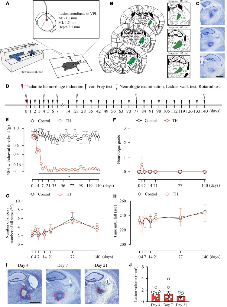
Figure 1. Thalamic hemorrhage induces mechanical allodynia without affecting motor functions.
(A) Schema of the thalamic hemorrhage (TH) induction method. VPL, ventral posterolateral nucleus; AP, anterior-posterior; ML, medial-lateral. (B) Anatomy of our injection target, the VPL, and its surrounding area. The ventrobasal complex (VB) consists of the VPL and ventral posteromedial nucleus (VPM). (C) Representative image of the Nissl-stained tissue sections. Scale bar: 1 mm. (D) An experimental design showing the time-course of TH induction and behavioral tests. (E) The 50% withdrawal threshold of the contralesional hind paw was significantly decreased 5 days after TH induction and persisted throughout the testing period (*P < 0.01, 2-way repeated measures ANOVA followed by Sidak’s multiple comparisons test). (F–H) No motor deficit was observed in TH mice. (F) The neurologic grade (P > 0.05, group effect by 2-way repeated measures ANOVA). (G) The ladder walk test (P > 0.05, group effect by 2-way repeated measures ANOVA). (H) The rotarod assessment (P > 0.05, group effect by 2-way repeated measures ANOVA). (I and J) There was no significant difference in the lesion area quantified on days 4, 7, and 21 after TH (P > 0.05, group effect by 1-way repeated measures ANOVA). Red outlines in images represent the edges of lesions. Scale bar: 1 mm.

