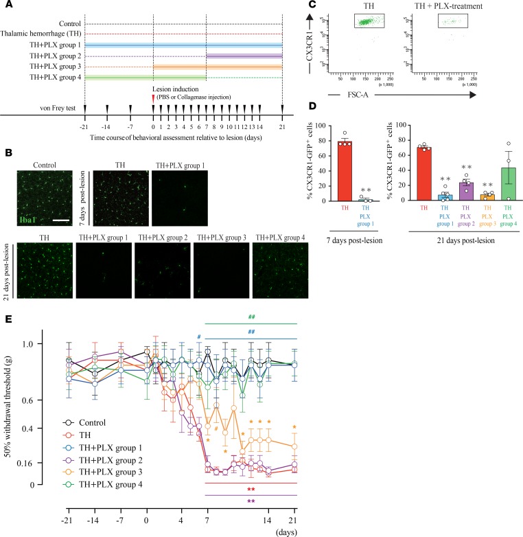
Figure 3. Microglial depletion prevented the development of mechanical allodynia induced by thalamic hemorrhage.
(A) Schematic of the experimental design. Control group mice received PBS injection into the ventral posterolateral nucleus (VPL) (n = 4). Thalamic hemorrhage (TH) group mice received collagenase injection and were fed control chow. TH + PLX group 1 mice were treated with PLX3397 from 21 days before hemorrhage until 21 days after hemorrhage (n = 4). TH + PLX group 2: from days 7–21 (n = 4). TH + PLX group 3: from days 0–21 (n = 4). TH + PLX group 4: from 21 days before to 7 days after TH (n = 5). (B) Iba-1 immunostaining of the S1 showed a robust decrease in microglial cells by PLX treatment. Scale bar: 100 μm. (C) Dot plots show the gating of CX3CR1 in each group. The plots were used to quantify the amount of microglia after TH. CX3CR1+/EGFP mice were used in the flow cytometry experiment. (D) Flow cytometry quantification showed that the number of microglia in the group treated with PLX3397 was significantly reduced compared with that in the TH group on day 7 (**P < 0.01, unpaired t test) and on day 21 (**P < 0.01, 1-way ANOVA followed by Dunnett’s multiple comparisons test) after hemorrhage. (E) The 50% withdrawal threshold in the TH group was significantly reduced after hemorrhage compared with the Control group (**P < 0.01, *P < 0.05, 2-way repeated measures ANOVA followed by Tukey’s multiple comparisons test). TH + PLX groups 1 and 4, which started PLX treatment before lesion induction, exhibited a higher withdrawal threshold than the TH group did (##P < 0.01, #P < 0.05, 2-way repeated-measures ANOVA followed by Tukey’s multiple comparisons test); however, TH + PLX group 2 and 3, which started PLX treatment after lesion induction, exhibited a lower withdrawal threshold, similar to the level exhibited by the TH group.

