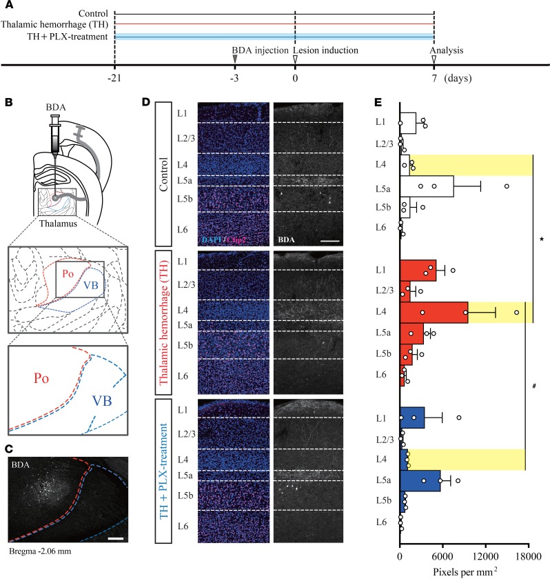
Figure 4. Thalamic hemorrhage induces abnormal formation of thalamocortical projections into layer 4 of the S1.
(A) Experimental design and timeline. (B and C) Biotinylated dextran amines (BDA) injection into the posterior nucleus (Po) of the thalamus adjacent to the VB. Scale bar: 200 μm. (D and E) Distribution of BDA-labeled Po axons in each cortical layer of the S1. The fluorescence intensity (pixel per mm2) was higher in cortical layer 4 of the thalamic hemorrhage (TH) group than in that of the TH + PLX group and Control group (*P < 0.01, layer 4 of TH group vs. layer 4 of Control group; #P < 0.01, layer 4 of TH group vs. layer 4 of TH + PLX group. 2-way ANOVA followed by Tukey’s multiple comparisons test. Scale bar: 200 μm.

