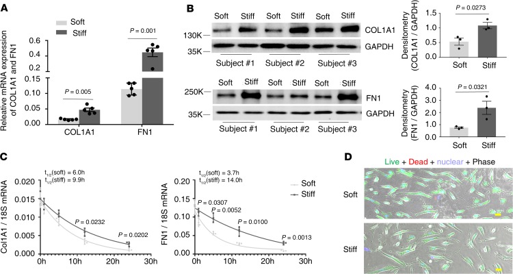
Figure 2. Stiff matrix promotes expression of COL1A1 and FN1 at both the mRNA and protein level.
Human lung fibroblast populations (n = 3) were cultured on soft and stiff polyacrylamide gels for 48 hours. (A) Relative levels of COL1A1 and FN1 mRNA were determined by real-time RT-PCR. GAPDH was used as internal reference control. Bar graphs represent (mean ± SD) 5 separate experiments. (B) Protein levels of COL1A1 and FN1 were determined by immunoblot. GAPDH was used as loading control. Relative density of COL1A1 and FN1 bands was normalized to GAPDH. Bar graphs represent (mean ± SD) 3 separate experiments. (C) Human lung fibroblasts were treated with 10 μg/mL actinomycin D, followed by culturing on soft and stiff polyacrylamide gels for 24 hours. Relative levels of COL1A1 and FN1 mRNA were determined by real-time RT-PCR at the indicated time points. 18S rRNA was used as internal reference control. Graphs represent mRNA decay and half-lives of COL1A1 and FN1 under soft and stiff matrix conditions after actinomycin D treatment and represent (mean ± SD) 3 separate experiments. (D) Cell viability after actinomycin D treatment for 24 hours under both soft and stiff matrix conditions was evaluated by live/dead cell viability assays. Scale bar: 20 μm. A 2-tailed Student’s t test was used for comparison between groups.

