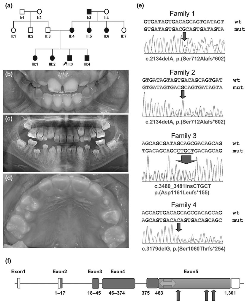
FIGURE 1.

Pedigree, clinical photograph, panoramic radiograph, sequencing chromatograms of family members and gene diagram of DSPP. (a) Pedigree of family 1. (b) Clinical photograph of the proband (III:3) at the age of 8 years. Remaining deciduous teeth show an amber-brown discoloration and mild to moderate attrition, but erupting permanent teeth look normal without any discoloration. (c) Panoramic radiograph of the proband at the age of 10 years reveals the characteristic thistle tube-shaped pulp chambers with pulp stones. (d) Clinical photograph of the proband of family 3 at the age of 9 years 8 months. Remaining deciduous teeth exhibit a dark brown discoloration and severe attrition. Permanent dentition is normal in shape and color in most teeth, but the lingual surfaces of the maxillary anterior teeth show a mild brown hue at the cervical area. (e) Sanger sequencing chromatograms of the mutations identified. Wild-type (wt) and mutant (mut) nucleotide sequences are written on the above each chromatogram. The location of the mutations (deletion and insertion) was indicated with a red arrow in each chromatogram. Sequences based on the reference sequence for mRNA (NM_014208.3), where the A of the ATG translation initiation codon is nucleotide 1. (f) DSPP consists of 5 exons. Boxes indicate exons, and the amino acid numbers encoded by the exon are shown below each exon. The white area indicates the non-coding part, and the blue area indicates DSP region. Orange color indicates DPP region (463-1301 amino acids). The area associated with DD-II is shown as a green double arrow. The red arrows indicate the position of identified mutations in this study
