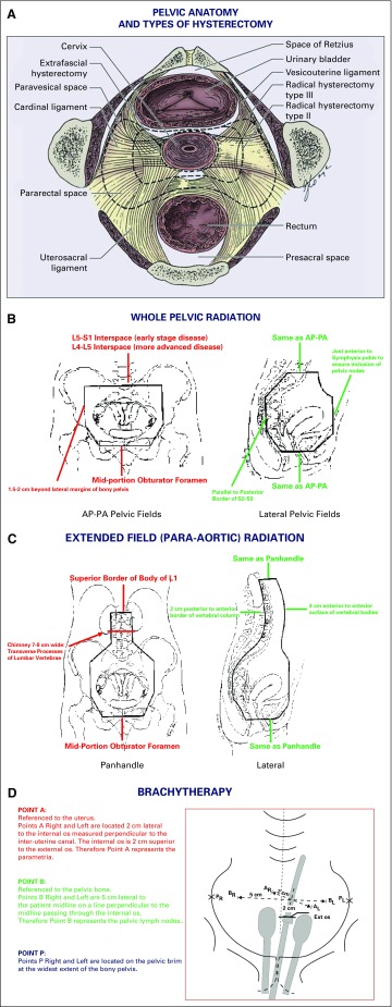
FIG 1.
Anatomic landmarks for surgical treatment of early stage cervical cancer (Panel A) and for administration of radiation therapy for locally advanced cervical cancer (Panels B-D). (A) Anatomy of the pelvis depicting location of the paravesical and pararectal spaces and other important anatomic landmarks encountered during performance of extrafascial, modified radical, and/or radical hysterectomy. (B) Wholepelvic radiotherapy. (C) Extended-field (para-aortic) radiotherapy. (D) Intracavitary brachytherapy. Point A: referenced to the uterus. Points A right (AR) and left (AL) are located 2 cm lateral to the internal os measured perpendicular to the interuterine canal. The internal os is 2 cm superior to the external (ext) os. Therefore, point A represents the parametria. Point B: referenced to the pelvic bone. Points B right (BR) and left (BL) are 5 cm lateral to the patient’s midline on a line perpendicular to the midline passing through the internal os. Therefore, point B represents the pelvic lymph nodes. Point P: points P right (PR) and left (PL) are located on the pelvic brim at the widest extent of the bony pelvis. AP, anteroposterior; L, lumbar vertebra; PA, postero-anterior; S, sacral vertebra. Source: (A) Public domain (Berek JS, Hacker NF); (B-D) Radiation therapy manual of the Gynecologic Oncology Group. Adapted and labeled by the authors. Used with permission through open-access granted by the National Cancer Institute.

