
Fig. 3.
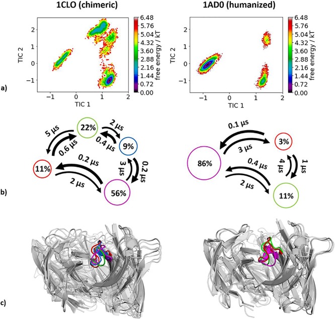
(a) Combined tICA plots of all available humanization variants using the same tICA coordinate system with the projected X-ray structures. AGless crystal structures are colored red. For the chimeric antibody we obtained 4.0 μs, for the humanized antibody 3.7 μs of molecular dynamics trajectories. (b) Markov-state models with the respective state probabilities and timescales. (c) Representative macrostate structures are shown and color-coded according to the state probabilities in (b).