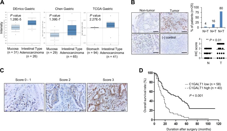
Fig. 1. C1GALT1 expression in gastric cancer.
a C1GALT1 mRNA expression in normal and cancerous gastric tissues in the Oncomine database. b C1GALT1 expression in paired gastric tumors. Immunohistochemical staining revealed C1GALT1 expression in paired gastric adenocarcinoma tumor (right) and nontumor mucosa tissue (left). In nontumor mucosa, foveolar epithelial cells (upper left) expressed less C1GALT1 than glandular epithelial cells (lower left). The negative control (lower right) did not exhibit specific staining. Scale bar, 50 µm. C1GALT1 was frequently overexpressed in gastric adenocarcinoma tumor (T) compared with its surrounding nontumor mucosa (N). *p < 0.05, paired t test. c Scoring of C1GALT1 expression (0–1, 2, and 3) analyzed using immunohistochemistry. Scale bar, 50 µm. d Kaplan–Meier survival analysis according to the expression of C1GALT1 in gastric cancer patients (n = 98).

