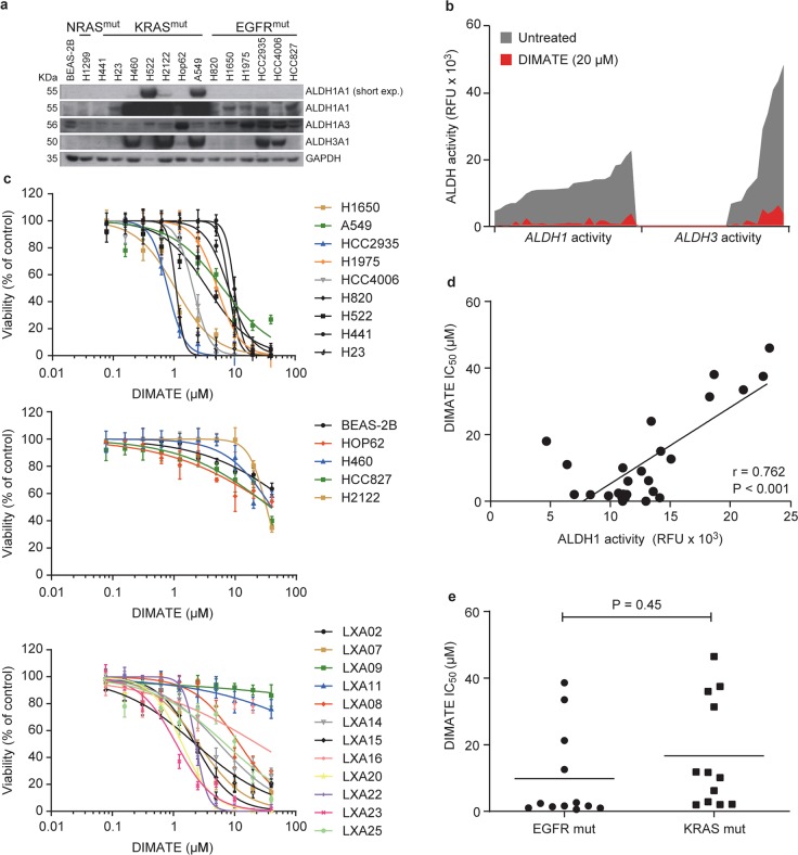
Fig. 2. DIMATE affects the viability of NSCLC cells independent of their genetic background.
a Immunoblots showing the amounts of ALDH1A1, ALDH1A3, and ALDH3A1 in normal human bronchial epithelial BEAS-2B cells and 14 NSCLC cell lines. GAPDH was used as the loading control. b Representative changes in ALDH1 and ALDH3 activity in an expanded panel of 26 NSCLC cell lines, including the cell lines in a and 12 xenograft-derived NSCLC primary cell lines (LXA), untreated or treated with the indicated dose of DIMATE. Data are plotted in increasing order according to the registered endogenous ALDH activity for each NSCLC cell line, i.e., from lower to higher mean values. A continuous connecting line was drawn to better illustrate the inhibition of the signal in the presence of DIMATE. ALDH activity was measured using a fluorometric enzymatic assay and two substrate probes (SEF0025 and SEF0013) with preferential affinity for ALDH class 1 and ALDH class 3 molecules, respectively (see the “Materials and methods” section for experimental details). c Dose-response curves for cell viability in the panel of 26 cell lines treated for 72 h with increasing doses of DIMATE. DIMATE-sensitive NSCLC cell lines are grouped in the upper plot; resistant cell lines, including the normal BEAS-2B line, are presented in the middle plot; and xenograft-derived NSCLC cells (LXA) are grouped in the lower plot. The error bars indicate the SDs (N = 4). d Graph showing the Pearson correlation between endogenous cellular ALDH1 activity and the IC50 values of DIMATE in the NSCLC cells in c. e Graphs showing the IC50 values of DIMATE in the NSCLC cell lines according to the mutational status. The horizontal bars indicate the mean values.

