FIGURE 2.
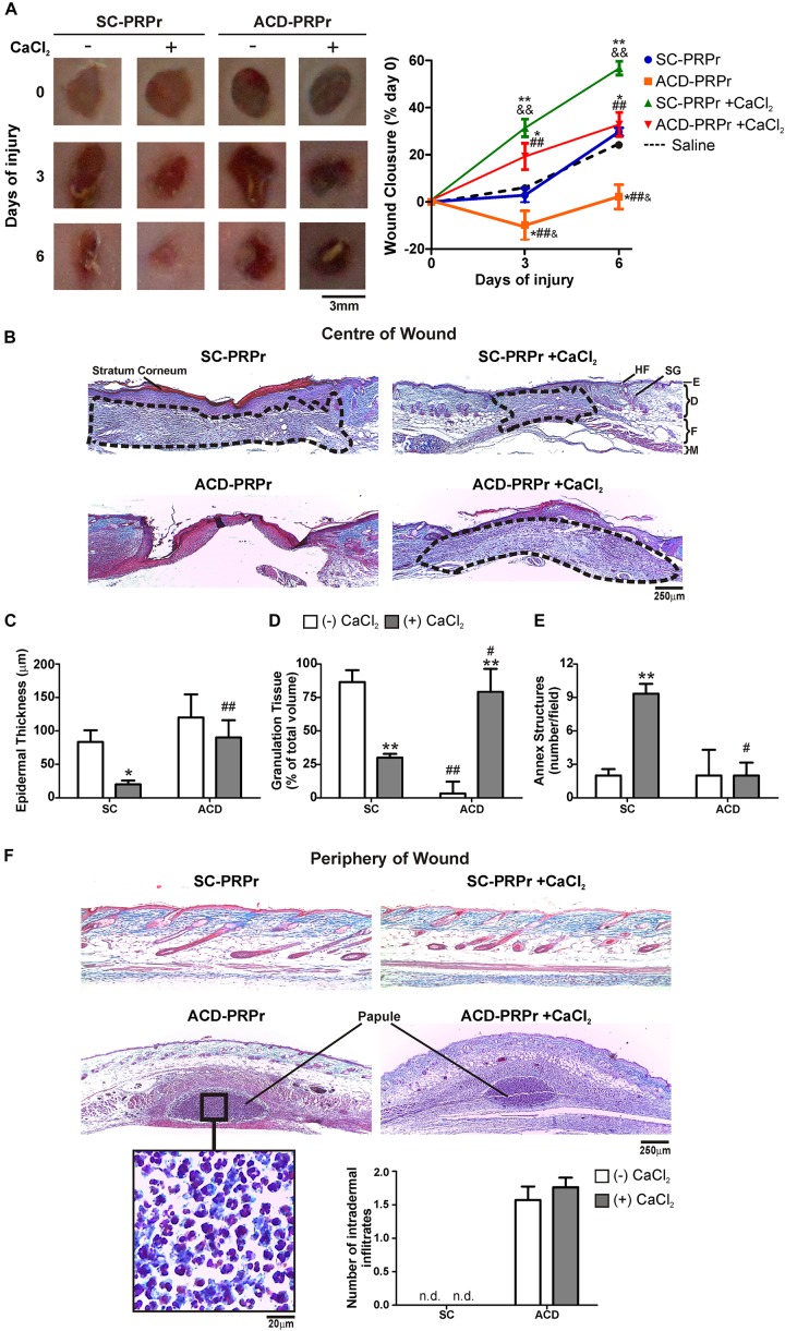
ACD but not SC induces inflammation and impairs PRPr-mediated mouse skin regeneration. Four round full-thickness excisional wounds of 3 mm in diameter were generated in the back skin of female BALB/c mice (8–10 weeks old). SC-PRPr or ACD- obtained from other mice were supplemented or not with CaCl2 and injected subcutaneously in the periphery of wounds. Saline was used as control. Healing was analyzed at 3 and 6 days post-injury. (A) Wounds were photographed and wound closure% was determined by quantification of wounds perimeters using the ImageJ software. (n = 14; &P < 0.05, &&P < 0.01 vs. saline; *P < 0.05; **P < 0.01 vs. CaCl2 (-); ##P < 0.01 vs. SC-PRPr. Repeated Measures One-way ANOVA followed by Fisher test). (B) Skin biopsies obtained on day 6 were stained with Masson’s trichrome. Images of the center of wounds were captured using an inverted microscope. (C) Epidermal thickness, (D) granulation tissue volume (dotted lines), and (E) annex structures (hair follicles and sebaceous glands) were quantified using the ImageJ software. (F) Images of the periphery of wounds were captured and intradermal inflammatory infiltrates were quantified. SG, sebaceous gland; HF, hair follicle; E, epidermis; D, dermis; F, fat layer; M, muscle layer. (Magnification 100X). (n = 14; *P < 0.05; **P < 0.01 vs. CaCl2 (-); #P < 0.05, ##P < 0.01 vs. SC-PRPr. Two-way ANOVA followed by Fisher test).

