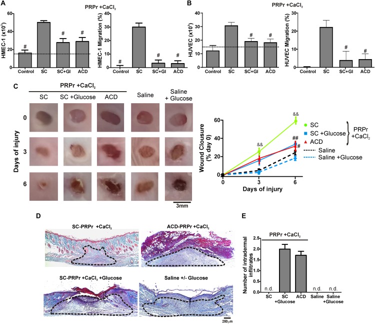
FIGURE 3.
D-Glucose (Dextrose) confers anti-angiogenic, anti-regenerative and inflammatory proprieties to PRP. HMEC-1 (A) or HUVEC (B) were incubated with SC-PRPr + CaCl2 (supplemented or not with Glucose 18 mM) or ACD-PRPr + CaCl2. MCDB-131 medium (A) or Endothelial Basal Medium 2 (EBM2) (B) both supplemented with FBS 2% were used as control. Endothelial cell proliferation at 24 h and migration at 8 h were determined (n = 4; #P < 0.05 vs. SC-PRPr. Kruskal-Wallis test followed by Dunn test). (C–E) Murine SC-PRPr + CaCl2 (supplemented or not with Glucose 18 mM) or ACD- were injected subcutaneously in the periphery of wounds generated in back skin of other mice. Saline supplemented or not with Glucose was used as control. Healing was analyzed at 3 and 6 days post-injury. (C) Wounds were photographed to determine the wound closure% (n = 8, &&P < 0.01 vs. saline; #P < 0.05, ##P < 0.01 vs. SC-PRPr. Repeated Measures One-way ANOVA followed by Fisher test). (D) Skin biopsies obtained on day 6 were stained with Masson’s trichrome. Images of the center of wounds were captured using an inverted microscope (Magnification 100X). (E) Number of intradermal inflammatory infiltrates in the periphery of wounds was quantified (n = 8).

