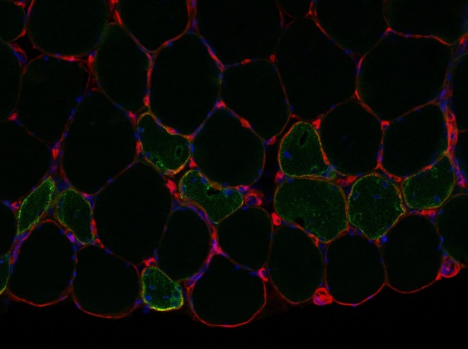
Figure 2.

Small moderately strongly to strongly NCAM-positive muscle fibers from a subject in the study of Bjørnsen et al. (2019). The biopsy was taken 3 days after the last training week. Note central/non-peripheral myonuclei in several of the NCAM-positive fibers. The section was re-photographed (due to loss of the original pictures) after several years in the freezer, and the positive staining would likely have been even stronger if the section was new. Red = laminin, green = NCAM, and blue = DAPI. Picture cropped from 10× original. Picture courtesy of Mathias Wernbom.
