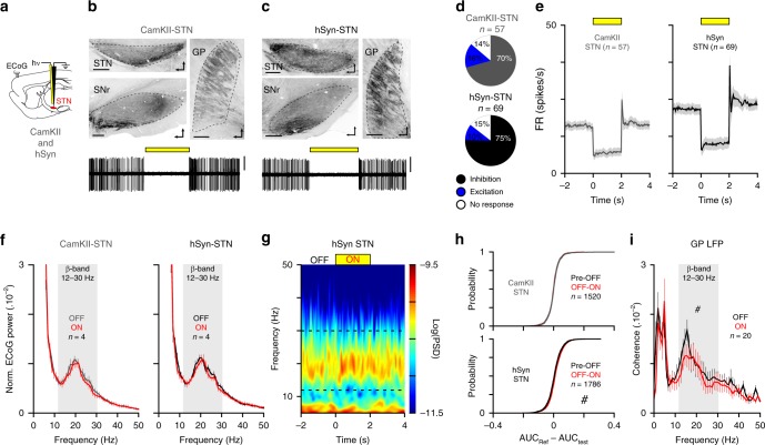
Fig. 2. STN activity is not necessary for abnormal β-synchronization in cortico-BG loop.
a Schematic of STN opto-inhibition using CamKII or a ubiquitous and neuron-specific promotor: the human synapsin 1 (hSyn) dependent virus in 6-OHDA rats. b, c Sagittal epifluorescence images (top) showing the EYFP expression in STN and the appropriate axonal labeling in GP and substantia nigra pars reticulata (SNr). Bottom traces show examples of STN neuron opto-inhibition in CamKII-STN (b) or hSyn-STN (c) conditions (slice orientation: dorso-rostral, scale: 400 µm, scales: 0.5 mV, 6 s recording). d Proportion of STN neurons inhibited (black), excited (blue), or non-modulated (white) by the laser stimulation in CamKII-STN and hSyn-STN. e Population PSTH of STN firing during opto-inhibition in CamKII-STN (bin: 50 ms) and hSyn-STN groups (bin: 50 ms). f Mean normalized power spectrum of mCx ECoG (analysis on the best β epochs recordings) during OFF and ON laser stimulations in CamKII-STN (left) and hSyn-STN (right). g Example of peri-event spectrogram of mCx ECoG recording during STN opto-inhibition in hSyn-STN rat. h Cumulative distribution function of the β-AUCs differences calculated for AUC(Pre–OFF) versus AUC(OFF–ON) for all ECoG β epochs in CamKII-STN (top) and hSyn-STN (bottom) conditions. i Mean coherence for ECoG versus GP LFP at β-frequency during OFF and ON laser stimulation in hSyn-STN rats. Group data are mean ± SEM. See also Supplementary Table 2 for statistical information.

