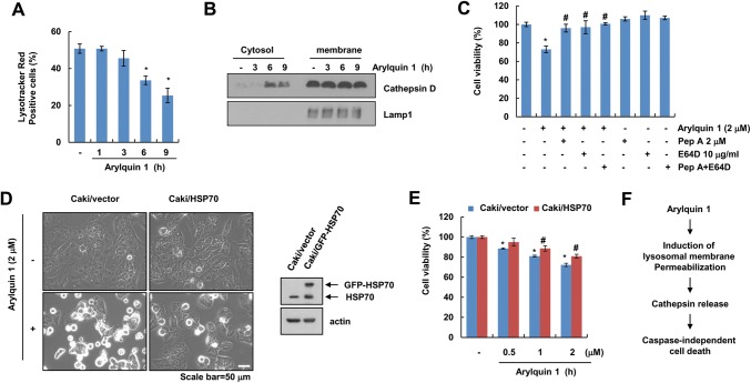
Fig. 4.
Arylquin 1 induces lysosomal membrane permeabilization-mediated cell death. a, b Caki cells were treated with 2 μM arylquin 1 for the indicated time periods, and then cells were incubated with the LysoTracker Red fluorescent dye. The fluorescence intensity was detected using flow cytometry (a). Cytosol and membrane fractions (lysosome-rich fraction) were prepared, and the protein expression levels of cathepsin D and Lamp1 were determined by western blotting (b). c Caki cells were pretreated with 2 μM pepstatin A (Pep A) and/or 10 μg/mL E64D for 30 min, and then added with 2 μM arylquin 1 for 24 h. Cell viability was determined using the XTT assay. d, e Caki/vector and Caki/HSP70 cells were treated with the indicated concentrations of arylquin 1 for 24 h. Cell morphology was examined using interference light microscopy (d). Cell viability was determined using the XTT assay (e). f Schematic diagram of arylquin 1-induced cell death in cancer cells. The protein expression levels of HSP70 and actin were determined by western blotting. The values in a, c, e represent the mean ± SEM from three independent samples. *p < 0.05 compared to the control. #p < 0.05 compared to the arylquin 1

