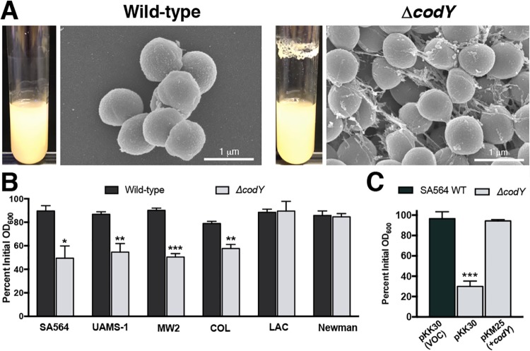
FIG 1.
ΔcodY mutant cells of diverse S. aureus clinical isolates form large cell aggregates tethered by a stringlike matrix. (A) Scanning electron microscopy was performed on SA564 and ΔcodY mutant cells during exponential growth in tryptic soy broth. Images are representative of multiple experiments. Images are at the same magnification. Representative images of biofilm observed in overnight culture growth are shown to the left of each micrograph. (B and C) Percent aggregation of S. aureus clinical isolates and their ΔcodY mutant derivatives (B) and the complemented SA564 codY-null mutant (C) using the settling assay from samples obtained during exponential growth in TSB as described in Materials and Methods. Data indicate the mean ± standard error of the mean (SEM) values from at least three independent experiments. *, P < 0.05; **, P < 0.01; ***, P < 0.001 (relative to WT using Student’s t test [B] and analysis of variance [ANOVA] with Dunnett’s postanalysis relative to SA564 WT [C]). VOC, vector-only control.

