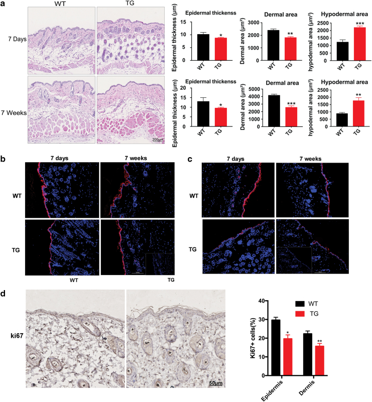
Figure 2.
Dermal skin thickness was reduced and hypodermal adipose tissue was increased in TG mice compared with WT controls. (a) Histological analysis of dorsal skin sections revealed that the dermal area of TG mice was reduced compared with WT controls 7 days after birth and persisted into adulthood. n = 6, *p < 0.05; **p < 0.01; ***p < 0.001 by two-tailed Student's t-test. (b) Immunofluorescence labeling of K10 in the skin of WT and TG mice (K10—red, DAPI—blue). Scale bar = 0.1 mm. (c) Immunofluorescence labeling of K14 in the skin of WT and TG mice (K14—red, DAPI—blue). Scale bar = 0.1 mm. (d) Representative immunohistochemical analyses of Ki67 (brown-stained nuclei in the epidermis and dermis) in the skin of WT and TG mice. Scale bar = 50 μm. *p < 0.05; **p < 0.01. DAPI, 4′,6-diamidino-2-phenylindole; TG, transgenic; WT, wild type. Color images are available online.

