Figure 3.
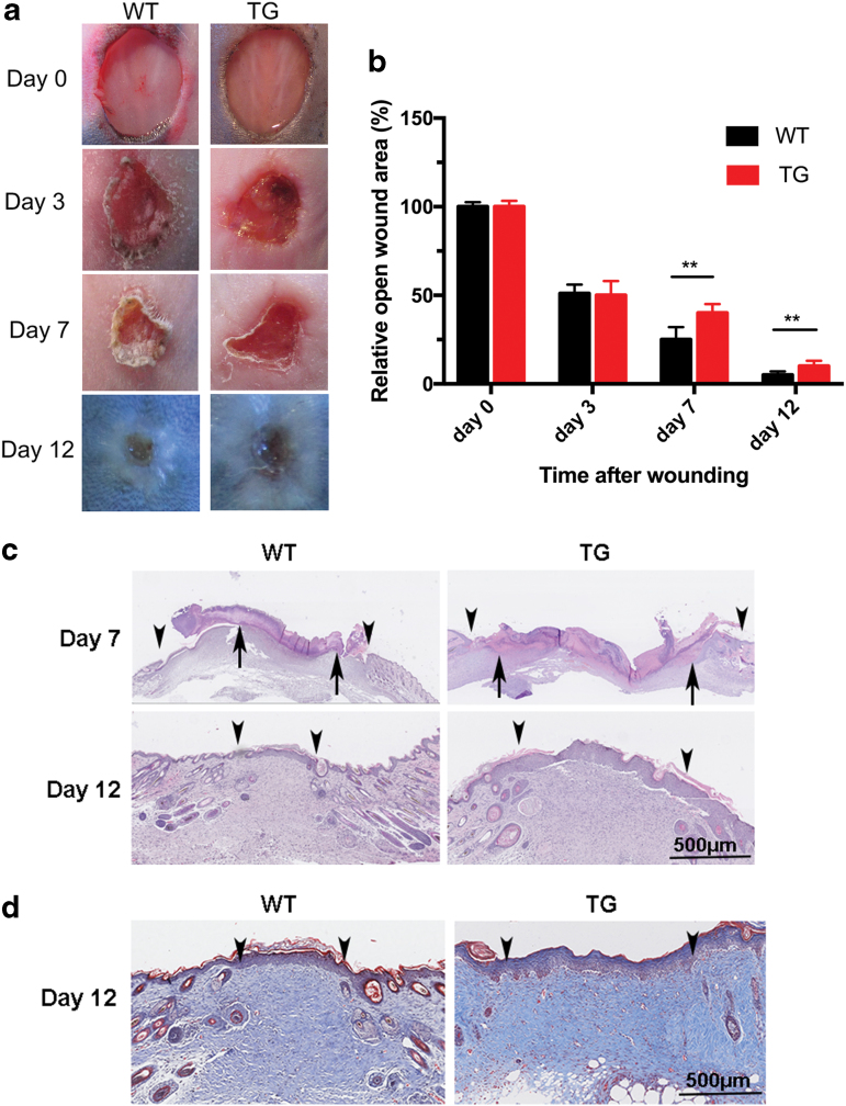
Cutaneous wound healing was delayed in TG mice. (a) Representative images of wounds generated with dermal punch biopsies on the dorsal skin of 7-week-old male mice show significant delays in wound closure in TG mice compared with WT controls. Duplicate wounds, n = 6, (b) the proportion of the wound remaining open relative to the initial wound area at each time point. Data are shown as mean ± standard error of the mean from six to eight wounds and are representative of three independent experiments with similar results. **p < 0.01, by two-tailed Student's t-test. (c) Hematoxylin and eosin staining of reepithelialization (wound margin [arrowheads] and the leading edge of epithelial wound [arrows]). Scale bar = 500 μm. (d) Masson's trichrome-stained wound sections at 12 days postinjury show the extent of granulation tissue at the midpoint of the wound (wound margin [arrowheads]). Scar bar = 500 μm. TG, transgenic; WT, wild type. Color images are available online.

