Figure 2.
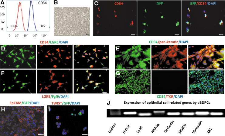
Enrichment and epithelial-like differentiation of CD34+ BDPCs. (A) After MACS, enriched CD34+ cells achieved near 100% purity in 10 days of expansion. (B) BDPCs cocultured with AML12 cells demonstrate the morphological changes, including increased volume, a flat and elongated shape, and a decreased nuclei to cytoplasm ratio. (C) Co-expression of CD34 and GFP confirms that the enriched cells retain GFP expression. (D) Immunofluorescent double staining demonstrates that enriched BDPCs co-express CD34 and LGR5. (E) The CD34+ cells express epithelial marker pan-keratin. (F) LGR5 positive cells synthesize and express Fgf9. (G) The epithelial-like differentiated BDPCs (eBDPCs) are negative for TCR antibody, a specific marker for γδ T lymphocytes. (H) Clear surface expression of epithelial cell marker EpCAM on the cellular membrane of cultured eBDPCs. (I) Twist expressed in eBDPCs. (J) PCR analysis reveals the active expression of epithelial transition-related genes, such as Notch, Snail, HNF4α, Occludin, MMP9, and Vimentin. Scale bar = 10 μm. EpCAM, epithelial cell adhesion molecule; Fgf9, fibroblast growth factor 9; HNF, hepatocyte nuclear factor; MACS, magnetic-activated cell sorting; TCR, T cell receptor.

