Figure 5.
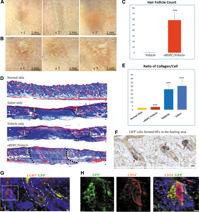
HFN post-eBDPC treatment. (A) The wound healing area of the vehicle-treated wounds (upper panel, v1–v3) healed with large or contracted scar region without hair formation. (B) The eBDPC-treated wound area (lower panel, e1–e3) demonstrates hair growing around the wound edge or in the center of the wound region. (C) HF count showed increased new HFN in the wound area on the histological section (***p < 0.001). (D) With trichrome staining, the normal skin of FVB mouse demonstrates an intact clearly layered cellular structures. Saline-treated wound healing shows a large amount of collagen mass lacking cellular structures filling the wound gap. Vehicle-treated wound also shows a large area of collagen deposit. The fibrin gel caused some inflammatory cell infiltration in the wound area. The eBDPC-treated mice show a reduction in fibrous tissue deposits, and a high density of HFs formed in the scar area. The disrupted subdermal adipose and smooth muscle tissues are used to define the edges of all wounds (yellow arrows and dotted line). (E) The graph shows a significant lower collagen deposit in the eBDPC treatment group (***p < 0.01). (F) IF staining shows GFP+ cells (green) in HFs within the wound healing area. (G) GFP+ cells co-express LGF5 (red) in the HFs of the wound area. (H) Two HFs are seen in the wound area. Both HFs are CD34+, but only the one on the left strongly expresses GFP indicating that the newly formed HF is from transplanted cells. Scale bar = 20 μm except for specifically labeled. FVB, Friend leukemia virus B; HFs, hair follicles; IF, immunofluorescence.

