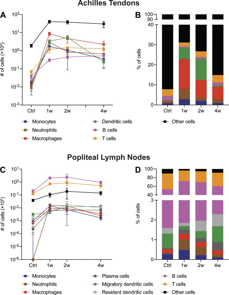
Fig. 6.
Summary of immune cell changes in Achilles tendons and lymph nodes after Achilles tenotomy and repair. The absolute number (A) and percentage (B) of monocytes, neutrophils, macrophages, dendritic cells, B cells, T cells, and other cells from Achilles tendons in control tendons and tendons 1 wk (1w), 2 wk (2w), or 4 wk (4w) after tenotomy and repair are shown. The absolute number (C) and percentage (D) of monocytes, neutrophils, macrophages, plasma cells, migratory dendritic cells, resident dendritic cells, B cells, T cells, and other cells from popliteal lymph nodes in control tendons and tendons 1w, 2w, or 4w after tenotomy and repair are shown.

