Fig. 2.
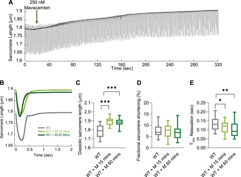
The effect of mavacamten on unloaded sarcomere shortening over time. Representative raw sarcomere length trace of a guinea pig left ventricular cardiomyocyte (GPCM) trace to show the real-time change to basal sarcomere length over time after addition of 250 nM mavacamten (green arrow; A). Averaged sarcomere length traces of wild-type (WT) control GPCMs ± mavacamten incubated for either 15 or 60 min (B). Diastolic sarcomere length (C), fractional shortening (D), and time to 50% (T50) relaxation (E) are plotted for WT, 250 nM mavacamten (M) for 15 min, and mavacamten (M) for 60 min; n = 30–41 cells from n = 4 isolations (B–E). Box and whisker plots (C–E) give the median average, interquartile range (box), and minimum and maximum data spread (whiskers). *P < 0.05, **P < 0.01, and ***P < 0.001.

