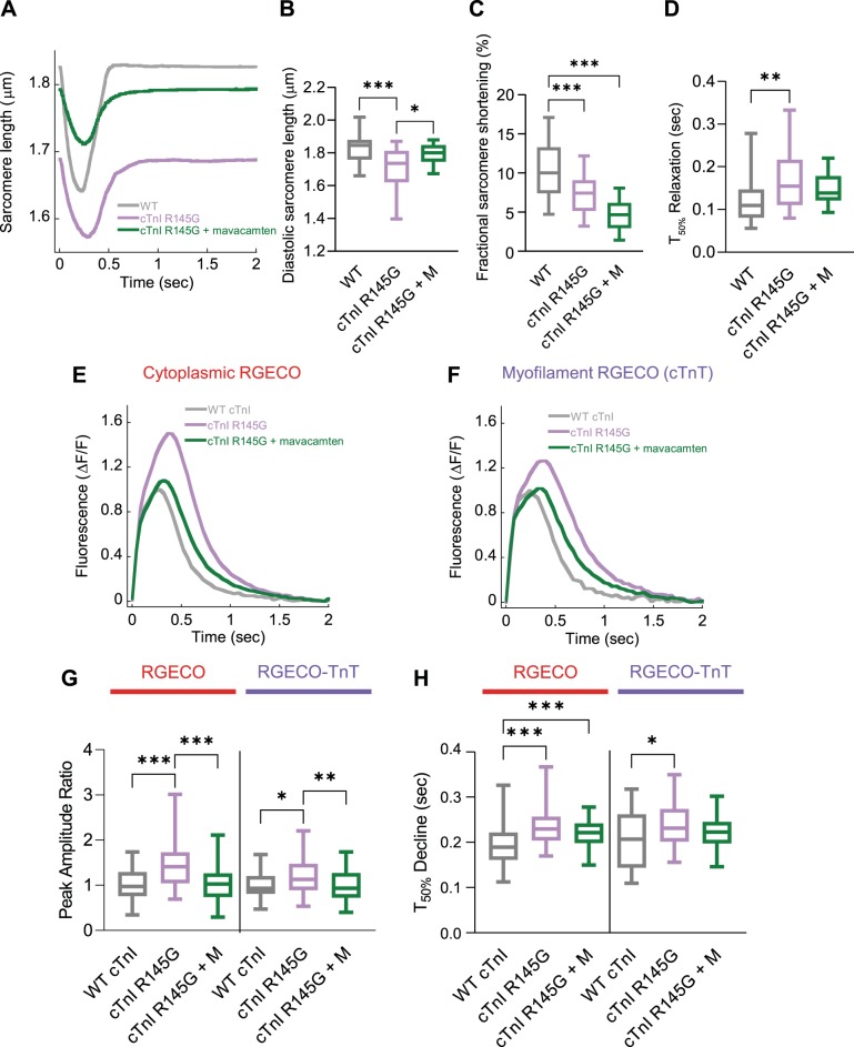
Fig. 4.
The effect of mavacamten on contractility and cytoplasmic/myofilament localized Ca2+ transients with adenovirally transduced cardiac troponin I (cTnI) R145G. Averaged sarcomere length traces of adult guinea pig left ventricular cardiomyocytes (GPCMs) transduced with either wild-type (WT) cTnI or cTnI R145G ± mavacamten (A). Diastolic sarcomere length (B), fractional shortening (C), and time to 50% (T50) relaxation (D) are plotted for WT, mutant troponin, and mutant troponin ± 250 nM mavacamten (M). Averaged Ca2+ transients GPCMs transduced with red-fluorescent, genetically encoded Ca2+ indicator for optical imaging (RGECO; E) or RGECO-TnT (F) and either WT cTnI or cTnI R145G ± 250 nM mavacamten. Peak amplitude ratios (G) and time to 50% decline (H) are plotted for WT, mutant troponin, and mutant troponin + 250 nM mavacamten (M); n = 12–32 (A–D) or n = 38–71 (E–H) cells from n = 3 isolations. Box and whisker plots (B–D, G, and H) give the median average, interquartile range (box), and minimum and maximum data spread (whiskers). *P < 0.05, **P < 0.01, and ***P < 0.001.

