Fig. 1.
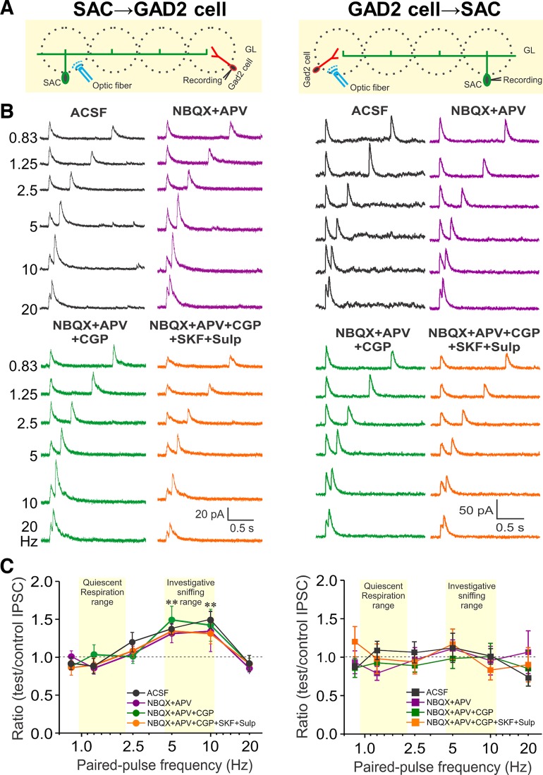
Differential short-term plasticity at reciprocal inhibitory synapses between short axon cells (SACs) and glutamic acid decarboxylase (GAD)2-expressing cells. A: schematic diagram showing the experimental design of optical stimulation of channelrhodopsin-2 (ChR2)-SACs (left) or GAD2 cells (right) and recording from GAD2 cells (left) or SACs (right). B: voltage-clamp recording from a GAD2 cell (left) or SAC (right) held at 0 mV in response to paired-pulse light stimulation at 6 different frequencies (0.83, 1.25, 2.5, 5, 10, and 20 Hz) in artificial cerebrospinal fluid (ACSF; black), 1,2,3,4-Tetrahydro-6-nitro-2,3-dioxo-benzo[f]quinoxaline-7-sulfonamide (NBQX) + d-2-amino-5-phosphonovalerate (APV) (purple), NBQX+APV+CGP55845 (CGP) (10 µM, green), and further addition of D1/D2 blockers [10 µM SKF83566 (SKF) and 100 µM sulpiride (Sulp)] (NBQX+APV+CGP+SKF+Sulp, orange). C: group data showing paired-pulse ratio of light-evoked inhibitory postsynaptic current (IPSC) peak amplitude of the test pulse to the control pulse at 6 different stimulation frequencies from 5 periglomerular cells and 8 SACs in ACSF and different treatments. **P < 0.01.

