Fig. 3.
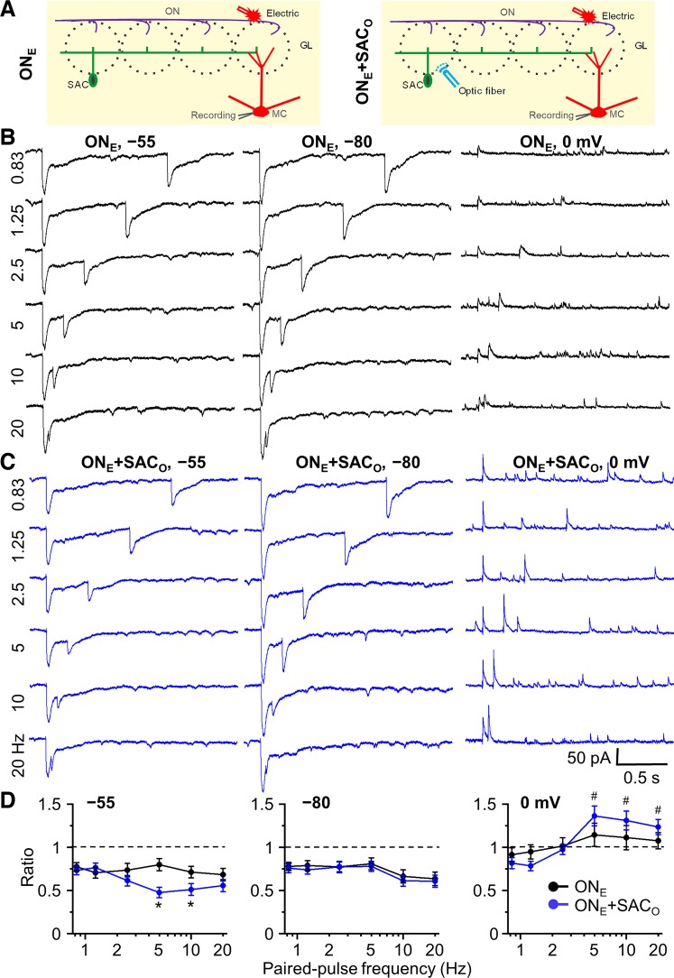
Impact of frequency-dependent short-term plasticity of short axon cell (SAC) inhibition on olfactory nerve electrical stimulation-induced synaptic currents in mitral cells (MCs). A: schematic diagram showing the experimental design of electric stimulation of olfactory nerve (ONE, left) and both ONE and optical stimulation of channelrhodopsin-2 (ChR2)-SACs (ONE+SACO, right). B: MC currents were recorded at 3 different holding potentials: −55 mV (~MC resting membrane potential; Liu and Shipley 2008), −80 mV [to isolate excitatory postsynaptic currents (EPSCs) with minimal Cl− driving force for inhibitory postsynaptic currents (IPSCs)], and 0 mV (to isolate IPSCs by enhancing Cl− driving force and minimizing EPSCs). Example traces of paired-pulse electric stimulation-induced EPSCs (inward current) at the holding potential of −55 and −80 mV, and IPSCs (outward current) at 0 mV with different paired-pulse frequencies of 0.83, 1.25, 2.5, 5, 10, and 20 Hz. C: example traces of ONE+SACO-induced EPSCs and IPSCs at different holding potentials with different paired-pulse frequencies. D: group data for paired-pulse ratio of postsynaptic currents of the test pulse to the control at the holding potentials of −55, −80, and 0 mV with different paired-pulse frequencies. ONE and ONE+SACO at different frequencies consistently induced paired-pulse depression of EPSCs at holding potential of −55 and −80 mV. ONE+SACO significantly increased paired-pulse depression at holding potential −55 mV at frequency of 5 and 10 Hz but did not enhance the depression at −80 mV at all frequencies. In contrast, at holding potential of 0 mV, ONE+SACO induced paired-pulse facilitation at 5, 10, and 20 Hz; ONE alone did not induce significant paired-pulse plasticity. The strength of optical and electric stimulation was adjusted to 50% maximal IPSC (at holding potential of 0 mV) and EPSC (−55 mV), respectively, so that either enhanced or suppressed IPSCs/EPSCs could be observed. Optical stimulation: 0.6–4 mW; electric stimulation: 15–45 µA. *P < 0.01 vs. ONE and #P < 0.01 vs. ratio of 1; n = 6. GL, glomerular layer.

