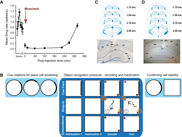
Fig. 1.
A: mean ± SE normalized firing frequency of CA1 hippocampal neurons before, during and at multiple time points after a local infusion of muscimol. Time 0 = the start of the infusion of muscimol. B: schematic depicting the in vivo recording and functional inactivation protocols. The darker blue background highlights the specific sessions for the three different object recognition (OR) protocols. Top: Cue Card OR, white square arena with dark cue card on one wall throughout all sessions. Middle: Drop-In OR, white square arena (no polarizing cue) where the mouse explored the empty arena for 2 min, after which the objects were placed into the arena in the positions indicated by the curved arrows. Bottom: conventional OR, same white square arena as in Drop-In OR, except that objects were present from the beginning upon mice entering the arena for the object sessions. Mice who received bilateral intrahippocampal infusion of saline or muscimol (i.e., inactivation studies) completed only the sessions in the darker blue section of the figure, while mice implanted with microdrive/tetrode arrays (i.e., in vivo recording studies) completed the lighter blue cue card rotations before, and immediately after, behavioral testing in the OR protocols (while place cells were recorded), to confirm cell stability and unit isolation. C, top: representative placement of the unilateral tetrode arrays implanted over the CA1 region of the right dorsal hippocampus for all mice (black dots). Bottom: representative photomicrograph of tetrode tip location in the dorsal hippocampus. D, top: representative bilateral intrahippocampal infusion sites within the CA1 region of the dorsal hippocampus for the Cue Card OR experiment (black dots). These sites are representative of the infusion locations for all of the inactivation experiments. Bottom: characteristic photomicrograph of the intrahippocampal microinfusion site into the CA1. Abbreviations in photomicrographs in C and D: CA1, CA2, and CA3, respective pyramidal cell fields of the hippocampus; DG, dentate gyrus; LPtA, lateral parietal association cortex; MPtA, medial parietal association cortex; RSA, retrosplenial agranular cortex; RSG, retrosplenial granular cortex; S1TR, primary somatosensory cortex trunk region.

