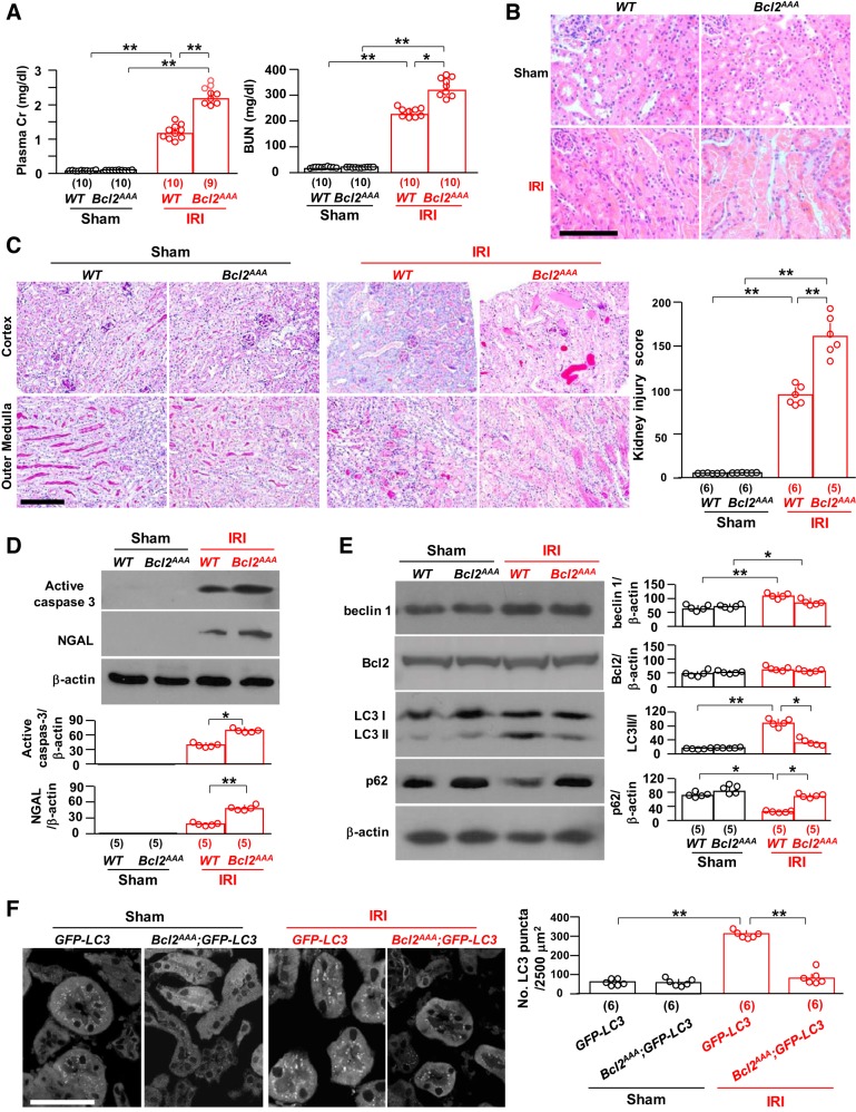
Fig. 4.
Loss of phosphorylation sites in Bcl-2 (Bcl2AAA) stabilizes the beclin 1/Bcl-2 complex and renders the kidney more susceptible to ischemia-reperfusion injury (IRI). In A–D, Bcl2AAA mice and their wild-type (WT) littermates were subjected to IRI or sham operations. Two days after surgery, mice were euthanized, blood was drawn, and kidneys were harvested. A: kidney function. Left, plasma creatinine (Cr); right, blood urea nitrogen (BUN). B: kidney histology with hematoxylin and eosin staining. Representative images are shown. Scale bar = 100 μm. C: kidney histology with periodic acid-Schiff (PAS) staining. Left, representative microscopic images of PAS staining. Scale bar = 100 μm. Right, semiquantitative assessment of kidney injury score based on PAS-stained sections in each group. D: kidney damage markers. Top, representative immunoblots for active caspase-3 and neutrophil gelatinase-associated lipocalin (NGAL) proteins in total kidney lysates. Bottom, summary of all immunoblots from each group. E: kidney autophagic markers. Left, representative immunoblots for beclin 1, Bcl-2, light chain (LC)3, and p62 in total kidney lysates. Right, summary of all immunoblots from each group. F: green fluorescent protein (GFP)-LC3 mice and Bcl2AAA;GFP-LC3 mice were subjected to IRI or sham operations. Two days after surgery, mice were euthanized, and kidneys were harvested. Left, representative microscopic images of GFP-LC3 punctas in the kidneys of GFP-LC3 mice and Bcl2AAA;GFP-LC3 mice by immunohistochemistry. Scale bar = 50 μm. Right, quantitative analysis of the number of GFP-LC3 puncta in renal tubules. Data are expressed as means ± SD, and statistical significance was evaluated by two-way ANOVA followed by a Student-Newman-Keuls post hoc test and significance was accepted when *P < 0.05 and **P < 0.01 between two groups. Sample numbers in each group are presented in brackets underneath the corresponding bars.

