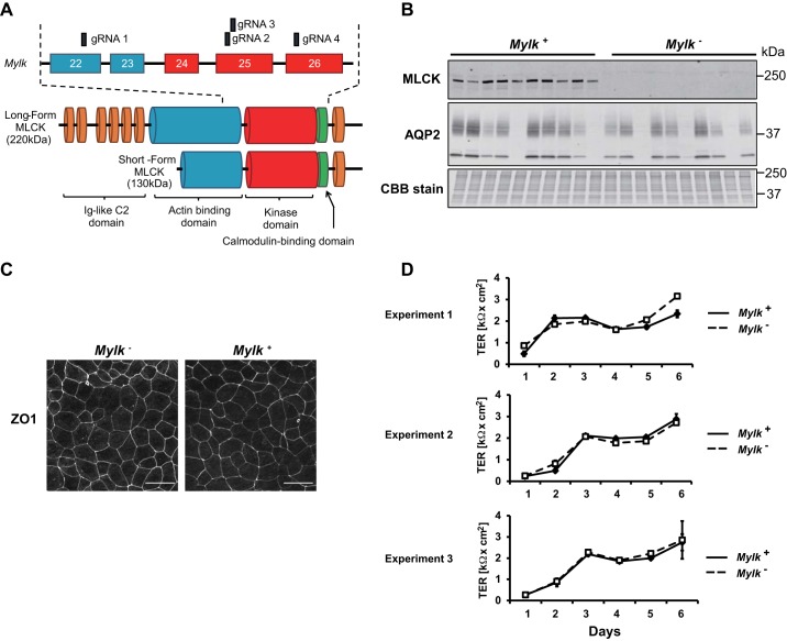
Fig. 2.
Generation and characterization of myosin light chain kinase (MLCK)-null and MLCK-intact cells. A: domain architecture of the Mylk gene and MLCK protein. Exons targeted by guide RNAs (gRNAs) are indicated. B: immunoblot of multiple MLCK-intact (Mylk+) and MLCK-null (Mylk−) clones showing MLCK protein levels along with aquaporin-2 (AQP2) protein levels and a Coomassie brilliant blue (CBB)-stained gel of total protein. C: MLCK-intact and MLCK-null cells formed polarized monolayers, as shown by zonula occludens 1 (ZO-1) staining, when grown on permeable membrane supports. D: development of transepithelial resistances (TERs) for three different pairs of MLCK-intact and MLCK-null cells.

