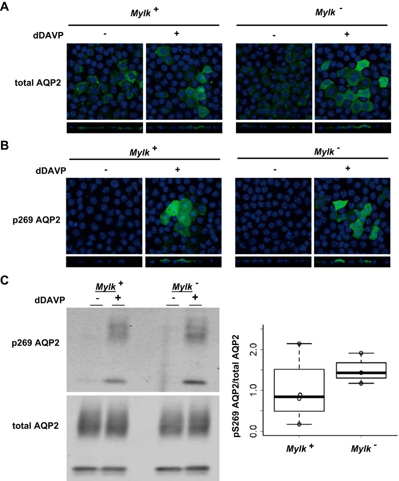
Fig. 3.
Apical distribution and phosphorylation of aquaporin-2 (AQP2) after exposure to 1-desamino-8-d-arginine-vasopressin (dDAVP). A and B: representative confocal images of myosin light chain kinase (MLCK)-intact (Mylk+) and MLCK-null (Mylk−) cells labeled with anti-AQP2 and anti-phosphorylated (S269) AQP2 (p269 AQP2) antibodies. Note redistribution of AQP2 from intracellular location to apical region in response to 30 min of exposure to 0.1 nM dDAVP. C: typical Western blot (left) and ratio of phosphorylated (S269) AQP2 to total AQP2 (right). Note the increased S269 phosphorylation of AQP2 after 30 min of treatment with dDAVP (0.1 nM) for both MLCK-intact and MLCK-null cells.

